1B9H | pdb_00001b9h

CRYSTAL STRUCTURE OF 3-AMINO-5-HYDROXYBENZOIC ACID (AHBA) SYNTHASE


Experimental Data Snapshot

  • Method: X-RAY DIFFRACTION
  • Resolution: 2.00 Å
  • R-Value Free: 
    0.252 (Depositor) 
  • R-Value Work: 
    0.215 (Depositor), 0.210 (DCC) 
  • R-Value Observed: 
    0.218 (Depositor) 

wwPDB Validation   3D Report Full Report


Ligand Structure Quality Assessment 

Created with Raphaël 2.3.0Worse 01 BetterLigand structure goodness of fit to experimental dataBest fitted PLPClick on this verticalbar to view details

This is version 1.3 of the entry. See complete history


Literature

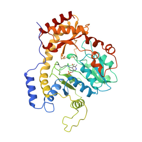
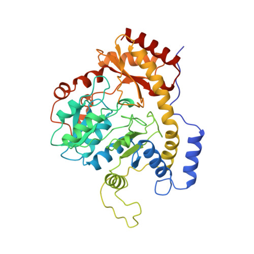
Crystal structure of 3-amino-5-hydroxybenzoic acid (AHBA) synthase.

Eads, J.C.Beeby, M.Scapin, G.Yu, T.W.Floss, H.G.

(1999) Biochemistry 38: 9840-9849

  • DOI: https://doi.org/10.1021/bi990018q
  • Primary Citation of Related Structures:  
    1B9H, 1B9I

  • PubMed Abstract: 

    The biosynthesis of ansamycin antibiotics, including rifamycin B, involves the synthesis of an aromatic precursor, 3-amino-5-hydroxybenzoic acid (AHBA), which serves as starter for the assembly of the antibiotics' polyketide backbone. The terminal enzyme of AHBA formation, AHBA synthase, is a dimeric, pyridoxal 5'-phosphate (PLP) dependent enzyme with pronounced sequence homology to a number of PLP enzymes involved in the biosynthesis of antibiotic sugar moieties. The structure of AHBA synthase from Amycolatopsis mediterranei has been determined to 2.0 A resolution, with bound cofactor, PLP, and in a complex with PLP and an inhibitor (gabaculine). The overall fold of AHBA synthase is similar to that of the aspartate aminotransferase family of PLP-dependent enzymes, with a large domain containing a seven-stranded beta-sheet surrounded by alpha-helices and a smaller domain consisting of a four-stranded antiparallel beta-sheet and four alpha-helices. The uninhibited form of the enzyme shows the cofactor covalently linked to Lys188 in an internal aldimine linkage. On binding the inhibitor, gabaculine, the internal aldimine linkage is broken, and a covalent bond is observed between the cofactor and inhibitor. The active site is composed of residues from two subunits of AHBA synthase, indicating that AHBA synthase is active as a dimer.


  • Organizational Affiliation

    School of Biochemistry, University of Birmingham, Edgbaston, Birmingham B15 2TT, U.K.


Macromolecules
Find similar proteins by:  (by identity cutoff)  |  3D Structure
Entity ID: 1
MoleculeChains Sequence LengthOrganismDetailsImage
PROTEIN (3-AMINO-5-HYDROXYBENZOIC ACID SYNTHASE)388Amycolatopsis mediterraneiMutation(s): 0 
EC: 4.2.1.144 (UniProt), 2.6.1 (UniProt)
UniProt
Find proteins for O52552 (Amycolatopsis mediterranei (strain S699))
Explore O52552 
Go to UniProtKB:  O52552
Entity Groups  
Sequence Clusters30% Identity50% Identity70% Identity90% Identity95% Identity100% Identity
UniProt GroupO52552
Sequence Annotations
Expand
  • Reference Sequence
Small Molecules
Ligands 1 Unique
IDChains Name / Formula / InChI Key2D Diagram3D Interactions
PLP
Query on PLP

Download Ideal Coordinates CCD File 
B [auth A]PYRIDOXAL-5'-PHOSPHATE
C8 H10 N O6 P
NGVDGCNFYWLIFO-UHFFFAOYSA-N
Experimental Data & Validation

Experimental Data

  • Method: X-RAY DIFFRACTION
  • Resolution: 2.00 Å
  • R-Value Free:  0.252 (Depositor) 
  • R-Value Work:  0.215 (Depositor), 0.210 (DCC) 
  • R-Value Observed: 0.218 (Depositor) 
Space Group: P 32 2 1
Unit Cell:
Length ( Å )Angle ( ˚ )
a = 89.7α = 90
b = 89.7β = 90
c = 127.7γ = 120
Software Package:
Software NamePurpose
SHARPphasing
REFMACrefinement

Structure Validation

View Full Validation Report


Ligand Structure Quality Assessment 

Created with Raphaël 2.3.0Worse 01 BetterLigand structure goodness of fit to experimental dataBest fitted PLPClick on this verticalbar to view details

Entry History 

Deposition Data

Revision History  (Full details and data files)

  • Version 1.0: 1999-08-13
    Type: Initial release
  • Version 1.1: 2008-04-26
    Changes: Version format compliance
  • Version 1.2: 2011-07-13
    Changes: Derived calculations, Version format compliance
  • Version 1.3: 2023-12-27
    Changes: Data collection, Database references, Derived calculations