An unexpected phosphate binding site in Glyceraldehyde 3-Phosphate Dehydrogenase: Crystal structures of apo, holo and ternary complex of Cryptosporidium parvum enzyme
Cook, W.J., Senkovich, O., Chattopadhyay, D.(2009) BMC Struct Biol 9: 9-9
- PubMed: 19243605
- DOI: https://doi.org/10.1186/1472-6807-9-9
- Primary Citation of Related Structures:
1VSU, 1VSV, 3CIF - PubMed Abstract:
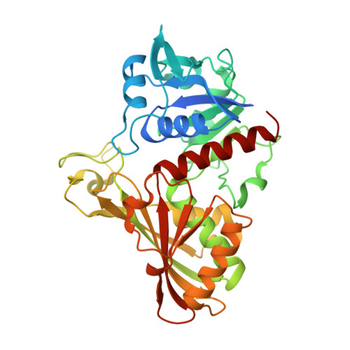
The structure, function and reaction mechanism of glyceraldehyde 3-phosphate dehydrogenase (GAPDH) have been extensively studied. Based on these studies, three anion binding sites have been identified, one 'Ps' site (for binding the C-3 phosphate of the substrate) and two sites, 'Pi' and 'new Pi', for inorganic phosphate. According to the original flip-flop model, the substrate phosphate group switches from the 'Pi' to the 'Ps' site during the multistep reaction. In light of the discovery of the 'new Pi' site, a modified flip-flop mechanism, in which the C-3 phosphate of the substrate binds to the 'new Pi' site and flips to the 'Ps' site before the hydride transfer, was proposed. An alternative model based on a number of structures of B. stearothermophilus GAPDH ternary complexes (non-covalent and thioacyl intermediate) proposes that in the ternary Michaelis complex the C-3 phosphate binds to the 'Ps' site and flips from the 'Ps' to the 'new Pi' site during or after the redox step. We determined the crystal structure of Cryptosporidium parvum GAPDH in the apo and holo (enzyme + NAD) state and the structure of the ternary enzyme-cofactor-substrate complex using an active site mutant enzyme. The C. parvum GAPDH complex was prepared by pre-incubating the enzyme with substrate and cofactor, thereby allowing free movement of the protein structure and substrate molecules during their initial encounter. Sulfate and phosphate ions were excluded from purification and crystallization steps. The quality of the electron density map at 2A resolution allowed unambiguous positioning of the substrate. In three subunits of the homotetramer the C-3 phosphate group of the non-covalently bound substrate is in the 'new Pi' site. A concomitant movement of the phosphate binding loop is observed in these three subunits. In the fourth subunit the C-3 phosphate occupies an unexpected site not seen before and the phosphate binding loop remains in the substrate-free conformation. Orientation of the substrate with respect to the active site histidine and serine (in the mutant enzyme) also varies in different subunits. The structures of the C. parvum GAPDH ternary complex and other GAPDH complexes demonstrate the plasticity of the substrate binding site. We propose that the active site of GAPDH can accommodate the substrate in multiple conformations at multiple locations during the initial encounter. However, the C-3 phosphate group clearly prefers the 'new Pi' site for initial binding in the active site.
Organizational Affiliation:
Department of Pathology, University of Alabama at Birmingham, Birmingham, AL 35294, USA. [email protected]


