2V5Z

Structure of human MAO B in complex with the selective inhibitor safinamide


Experimental Data Snapshot

  • Method: X-RAY DIFFRACTION
  • Resolution: 1.60 Å
  • R-Value Free: 
    0.227 (Depositor), 0.230 (DCC) 
  • R-Value Work: 
    0.208 (Depositor), 0.210 (DCC) 
  • R-Value Observed: 
    0.208 (Depositor) 

Starting Model: experimental
View more details

wwPDB Validation   3D Report Full Report


Ligand Structure Quality Assessment 

Created with Raphaël 2.3.0Worse 01 BetterLigand structure goodness of fit to experimental dataBest fitted FADClick on this verticalbar to view detailsBest fitted SAGClick on this verticalbar to view details

This is version 2.1 of the entry. See complete history


Literature

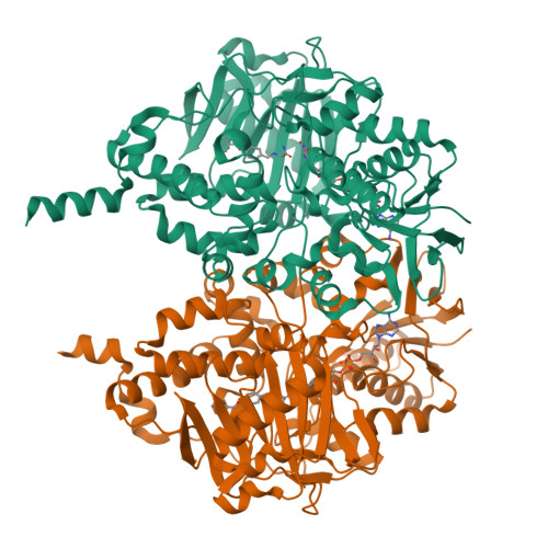
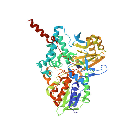
Structures of human monoamine oxidase B complexes with selective noncovalent inhibitors: safinamide and coumarin analogs.

Binda, C.Wang, J.Pisani, L.Caccia, C.Carotti, A.Salvati, P.Edmondson, D.E.Mattevi, A.

(2007) J Med Chem 50: 5848-5852

  • DOI: https://doi.org/10.1021/jm070677y
  • Primary Citation of Related Structures:  
    2V5Z, 2V60, 2V61

  • PubMed Abstract: 

    Structures of human monoamine oxidase B (MAO B) in complex with safinamide and two coumarin derivatives, all sharing a common benzyloxy substituent, were determined by X-ray crystallography. These compounds competitively inhibit MAO B with Ki values in the 0.1-0.5 microM range that are 30-700-fold lower than those observed with MAO A. The inhibitors bind noncovalently to MAO B, occupying both the entrance and the substrate cavities and showing a similarly oriented benzyloxy substituent.


  • Organizational Affiliation

    Department of Genetics and Microbiology, University of Pavia, via Ferrata 1, Pavia, 27100 Italy.


Macromolecules
Find similar proteins by:  (by identity cutoff)  |  3D Structure
Entity ID: 1
MoleculeChains Sequence LengthOrganismDetailsImage
Amine oxidase [flavin-containing] B
A, B
520Homo sapiensMutation(s): 0 
Gene Names: MAOB
EC: 1.4.3.4 (PDB Primary Data), 1.4.3.21 (UniProt)
UniProt & NIH Common Fund Data Resources
Find proteins for P27338 (Homo sapiens)
Explore P27338 
Go to UniProtKB:  P27338
PHAROS:  P27338
GTEx:  ENSG00000069535 
Entity Groups  
Sequence Clusters30% Identity50% Identity70% Identity90% Identity95% Identity100% Identity
UniProt GroupP27338
Sequence Annotations
Expand
  • Reference Sequence
Experimental Data & Validation

Experimental Data

  • Method: X-RAY DIFFRACTION
  • Resolution: 1.60 Å
  • R-Value Free:  0.227 (Depositor), 0.230 (DCC) 
  • R-Value Work:  0.208 (Depositor), 0.210 (DCC) 
  • R-Value Observed: 0.208 (Depositor) 
Space Group: C 2 2 2
Unit Cell:
Length ( Å )Angle ( ˚ )
a = 132.551α = 90
b = 223.602β = 90
c = 86.589γ = 90
Software Package:
Software NamePurpose
REFMACrefinement
MOSFLMdata reduction
SCALAdata scaling

Structure Validation

View Full Validation Report


Ligand Structure Quality Assessment 

Created with Raphaël 2.3.0Worse 01 BetterLigand structure goodness of fit to experimental dataBest fitted FADClick on this verticalbar to view detailsBest fitted SAGClick on this verticalbar to view details

Entry History 

Deposition Data

Revision History  (Full details and data files)

  • Version 1.0: 2007-10-16
    Type: Initial release
  • Version 1.1: 2011-05-08
    Changes: Version format compliance
  • Version 1.2: 2011-07-13
    Changes: Version format compliance
  • Version 2.0: 2023-01-18
    Type: Coordinate replacement
    Changes: Atomic model, Data collection, Database references, Derived calculations, Other, Refinement description, Source and taxonomy, Structure summary
  • Version 2.1: 2024-02-07
    Changes: Data collection, Refinement description