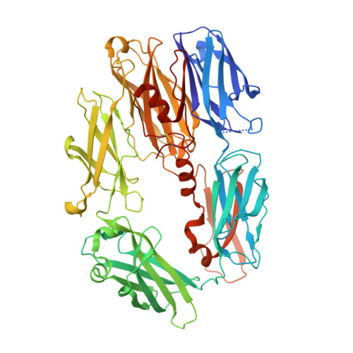
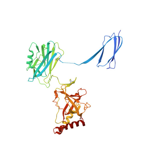
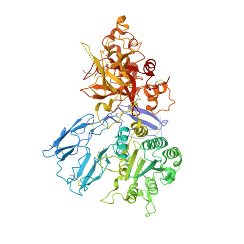
3HS0

Cobra Venom Factor (CVF) in complex with human factor B


Experimental Data Snapshot

  • Method: X-RAY DIFFRACTION
  • Resolution: 3.00 Å
  • R-Value Free: 0.243 
  • R-Value Work: 0.189 
  • R-Value Observed: 0.190 

Starting Model: experimental
View more details

wwPDB Validation   3D Report Full Report


Ligand Structure Quality Assessment 


This is version 2.4 of the entry. See complete history


Literature

Insights into complement convertase formation based on the structure of the factor B-cobra venom factor complex

Janssen, B.J.Gomes, L.Koning, R.I.Svergun, D.I.Koster, A.J.Fritzinger, D.C.Vogel, C.W.Gros, P.

(2009) EMBO J 28: 2469-2478

  • DOI: https://doi.org/10.1038/emboj.2009.184
  • Primary Citation of Related Structures:  
    3HRZ, 3HS0

  • PubMed Abstract: 

    Immune protection by the complement system critically depends on assembly of C3 convertases on the surface of pathogens and altered host cells. These short-lived protease complexes are formed through pro-convertases, which for the alternative pathway consist of the complement component C3b and the pro-enzyme factor B (FB). Here, we present the crystal structure at 2.2-A resolution, small-angle X-ray scattering and electron microscopy (EM) data of the pro-convertase formed by human FB and cobra venom factor (CVF), a potent homologue of C3b that generates more stable convertases. FB is loaded onto CVF through its pro-peptide Ba segment by specific contacts, which explain the specificity for the homologous C3b over the native C3 and inactive products iC3b and C3c. The protease segment Bb binds the carboxy terminus of CVF through the metal-ion dependent adhesion site of the Von Willebrand factor A-type domain. A possible dynamic equilibrium between a 'loading' and 'activation' state of the pro-convertase may explain the observed difference between the crystal structure of CVFB and the EM structure of C3bB. These insights into formation of convertases provide a basis for further development of complement therapeutics.


  • Organizational Affiliation

    Crystal and Structural Chemistry, Bijvoet Center for Biomolecular Research, Department of Chemistry, Faculty of Science, Utrecht University, Utrecht, The Netherlands.


Macromolecules
Find similar proteins by:  (by identity cutoff)  |  3D Structure
Entity ID: 1
MoleculeChains Sequence LengthOrganismDetailsImage
Cobra venom factorA,
E [auth F]
627Naja kaouthiaMutation(s): 0 
UniProt
Find proteins for Q91132 (Naja kaouthia)
Explore Q91132 
Go to UniProtKB:  Q91132
Entity Groups  
Sequence Clusters30% Identity50% Identity70% Identity90% Identity95% Identity100% Identity
UniProt GroupQ91132
Glycosylation
Glycosylation Sites: 1
Sequence Annotations
Expand
  • Reference Sequence
Find similar proteins by:  (by identity cutoff)  |  3D Structure
Entity ID: 2
MoleculeChains Sequence LengthOrganismDetailsImage
Cobra venom factorB,
F [auth G]
252Naja kaouthiaMutation(s): 0 
UniProt
Find proteins for Q91132 (Naja kaouthia)
Explore Q91132 
Go to UniProtKB:  Q91132
Entity Groups  
Sequence Clusters30% Identity50% Identity70% Identity90% Identity95% Identity100% Identity
UniProt GroupQ91132
Sequence Annotations
Expand
  • Reference Sequence
Find similar proteins by:  (by identity cutoff)  |  3D Structure
Entity ID: 3
MoleculeChains Sequence LengthOrganismDetailsImage
Cobra venom factorC,
G [auth H]
379Naja kaouthiaMutation(s): 0 
UniProt
Find proteins for Q91132 (Naja kaouthia)
Explore Q91132 
Go to UniProtKB:  Q91132
Entity Groups  
Sequence Clusters30% Identity50% Identity70% Identity90% Identity95% Identity100% Identity
UniProt GroupQ91132
Glycosylation
Glycosylation Sites: 1
Sequence Annotations
Expand
  • Reference Sequence
Find similar proteins by:  (by identity cutoff)  |  3D Structure
Entity ID: 4
MoleculeChains Sequence LengthOrganismDetailsImage
Complement factor BD,
H [auth I]
741Homo sapiensMutation(s): 2 
Gene Names: CFBBFBFD
EC: 3.4.21.47
UniProt & NIH Common Fund Data Resources
Find proteins for P00751 (Homo sapiens)
Explore P00751 
Go to UniProtKB:  P00751
PHAROS:  P00751
GTEx:  ENSG00000243649 
Entity Groups  
Sequence Clusters30% Identity50% Identity70% Identity90% Identity95% Identity100% Identity
UniProt GroupP00751
Glycosylation
Glycosylation Sites: 2Go to GlyGen: P00751-1
Sequence Annotations
Expand
  • Reference Sequence
Oligosaccharides

Help

Entity ID: 5
MoleculeChains Length2D Diagram Glycosylation3D Interactions
2-acetamido-2-deoxy-beta-D-glucopyranose-(1-4)-2-acetamido-2-deoxy-beta-D-glucopyranoseI [auth E],
J
2N-Glycosylation
Glycosylation Resources
GlyTouCan:  G42666HT
GlyCosmos:  G42666HT
GlyGen:  G42666HT
Small Molecules
Ligands 2 Unique
IDChains Name / Formula / InChI Key2D Diagram3D Interactions
NAG
Query on NAG

Download Ideal Coordinates CCD File 
K [auth A]
M [auth C]
N [auth D]
O [auth D]
Q [auth F]
K [auth A],
M [auth C],
N [auth D],
O [auth D],
Q [auth F],
S [auth H],
T [auth I]
2-acetamido-2-deoxy-beta-D-glucopyranose
C8 H15 N O6
OVRNDRQMDRJTHS-FMDGEEDCSA-N
MG
Query on MG

Download Ideal Coordinates CCD File 
L [auth A],
P [auth D],
R [auth F],
U [auth I]
MAGNESIUM ION
Mg
JLVVSXFLKOJNIY-UHFFFAOYSA-N
Experimental Data & Validation

Experimental Data

  • Method: X-RAY DIFFRACTION
  • Resolution: 3.00 Å
  • R-Value Free: 0.243 
  • R-Value Work: 0.189 
  • R-Value Observed: 0.190 
  • Space Group: P 21 21 21
Unit Cell:
Length ( Å )Angle ( ˚ )
a = 134.026α = 90
b = 136.975β = 90
c = 283.74γ = 90
Software Package:
Software NamePurpose
MOSFLMdata reduction
SCALAdata scaling
PHASERphasing
PHENIXrefinement
PDB_EXTRACTdata extraction
DNAdata collection

Structure Validation

View Full Validation Report


Ligand Structure Quality Assessment 


Entry History 

Deposition Data

Revision History  (Full details and data files)

  • Version 1.0: 2009-07-07
    Type: Initial release
  • Version 1.1: 2011-07-13
    Changes: Non-polymer description, Version format compliance
  • Version 2.0: 2020-07-29
    Type: Remediation
    Reason: Carbohydrate remediation
    Changes: Advisory, Atomic model, Data collection, Database references, Derived calculations, Structure summary
  • Version 2.1: 2021-03-31
    Changes: Source and taxonomy, Structure summary
  • Version 2.2: 2021-10-13
    Changes: Database references
  • Version 2.3: 2023-09-06
    Changes: Data collection, Refinement description
  • Version 2.4: 2024-10-30
    Changes: Structure summary