3W6U | pdb_00003w6u

Crystal structure of NADP bound L-serine 3-dehydrogenase from Hyperthermophilic Archaeon Pyrobaculum calidifontis


Experimental Data Snapshot

  • Method: X-RAY DIFFRACTION
  • Resolution: 2.00 Å
  • R-Value Free: 
    0.242 (Depositor), 0.217 (DCC) 
  • R-Value Work: 
    0.227 (Depositor), 0.207 (DCC) 

wwPDB Validation   3D Report Full Report


Ligand Structure Quality Assessment 

Created with Raphaël 2.3.0Worse 01 BetterLigand structure goodness of fit to experimental dataBest fitted NAPClick on this verticalbar to view details

This is version 1.2 of the entry. See complete history


Literature

Crystal structure of the NADP+and tartrate-bound complex of L-serine 3-dehydrogenase from the hyperthermophilic archaeon Pyrobaculum calidifontis.

Yoneda, K.Sakuraba, H.Araki, T.Ohshima, T.

(2018) Extremophiles 22: 395-405

  • DOI: https://doi.org/10.1007/s00792-018-1004-0
  • Primary Citation of Related Structures:  
    3W6U, 3W6Z, 3WS7, 5XVH

  • PubMed Abstract: 

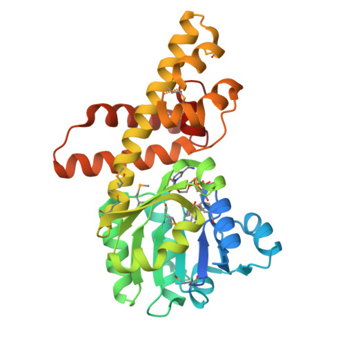
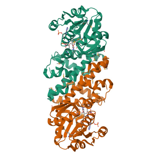
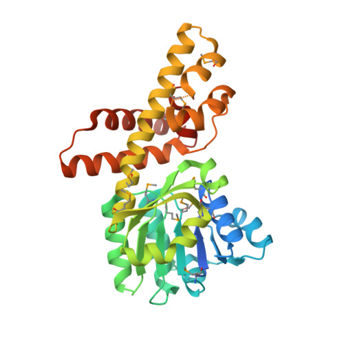
    A gene encoding L-serine dehydrogenase (L-SerDH) that exhibits extremely low sequence identity to the Agrobacterium tumefaciens L-SerDH was identified in the hyperthermophilic archaeon Pyrobaculum calidifontis. The predicted amino acid sequence showed 36% identity with that of Pseudomonas aeruginosa L-SerDH, suggesting that P. calidifontis L-SerDH is a novel type of L-SerDH, like Ps. aeruginosa L-SerDH. The overexpressed enzyme appears to be the most thermostable L-SerDH described to date, and no loss of activity was observed by incubation for 30 min at temperatures up to 100 °C. The enzyme showed substantial reactivity towards D-serine, in addition to L-serine. Two different crystal structures of P. calidifontis L-SerDH were determined using the Se-MAD and MR method: the structure in complex with NADP + /sulfate ion at 1.18 Å and the structure in complex with NADP + /L-tartrate (substrate analog) at 1.57 Å. The fold of the catalytic domain showed similarity with that of Ps. aeruginosa L-SerDH. However, the active site structure significantly differed between the two enzymes. Based on the structure of the tartrate, L- and D-serine and 3-hydroxypropionate molecules were modeled into the active site and the substrate binding modes were estimated. A structural comparison suggests that the wide cavity at the substrate binding site is likely responsible for the high reactivity of the enzyme toward both L- and D-serine enantiomers. This is the first description of the structure of the novel type of L-SerDH with bound NADP + and substrate analog, and it provides new insight into the substrate binding mechanism of L-SerDH. The results obtained here may be very informative for the creation of L- or D-serine-specific SerDH by protein engineering.


  • Organizational Affiliation

    Department of Bioscience, School of Agriculture, Tokai University, 9-1-1 Toroku, Higashi-ku, Kumamoto, Kumamoto, 862-8652, Japan. kyoneda@agri.u-tokai.ac.jp.


Macromolecules
Find similar proteins by:  (by identity cutoff)  |  3D Structure
Entity ID: 1
MoleculeChains Sequence LengthOrganismDetailsImage
6-phosphogluconate dehydrogenase, NAD-binding protein306Pyrobaculum calidifontis JCM 11548Mutation(s): 0 
Gene Names: Pcal_0699
EC: 1.1.1.276
UniProt
Find proteins for A3MU08 (Pyrobaculum calidifontis (strain DSM 21063 / JCM 11548 / VA1))
Explore A3MU08 
Go to UniProtKB:  A3MU08
Entity Groups  
Sequence Clusters30% Identity50% Identity70% Identity90% Identity95% Identity100% Identity
UniProt GroupA3MU08
Sequence Annotations
Expand
  • Reference Sequence
Small Molecules
Ligands 1 Unique
IDChains Name / Formula / InChI Key2D Diagram3D Interactions
NAP
Query on NAP

Download Ideal Coordinates CCD File 
B [auth A]NADP NICOTINAMIDE-ADENINE-DINUCLEOTIDE PHOSPHATE
C21 H28 N7 O17 P3
XJLXINKUBYWONI-NNYOXOHSSA-N
Modified Residues  1 Unique
IDChains TypeFormula2D DiagramParent
MSE
Query on MSE
A
L-PEPTIDE LINKINGC5 H11 N O2 SeMET
Experimental Data & Validation

Experimental Data

  • Method: X-RAY DIFFRACTION
  • Resolution: 2.00 Å
  • R-Value Free:  0.242 (Depositor), 0.217 (DCC) 
  • R-Value Work:  0.227 (Depositor), 0.207 (DCC) 
Space Group: C 1 2 1
Unit Cell:
Length ( Å )Angle ( ˚ )
a = 121.603α = 90
b = 56.827β = 106.69
c = 56.571γ = 90
Software Package:
Software NamePurpose
HKL-2000data collection
SOLVEphasing
CNSrefinement
HKL-2000data reduction
HKL-2000data scaling

Structure Validation

View Full Validation Report


Ligand Structure Quality Assessment 

Created with Raphaël 2.3.0Worse 01 BetterLigand structure goodness of fit to experimental dataBest fitted NAPClick on this verticalbar to view details

Entry History 

Deposition Data

Revision History  (Full details and data files)

  • Version 1.0: 2014-01-15
    Type: Initial release
  • Version 1.1: 2019-05-15
    Changes: Data collection, Database references, Derived calculations
  • Version 1.2: 2024-11-13
    Changes: Data collection, Database references, Derived calculations, Structure summary