3ZXS | pdb_00003zxs

Cryptochrome B from Rhodobacter sphaeroides


Experimental Data Snapshot

  • Method: X-RAY DIFFRACTION
  • Resolution: 2.70 Å
  • R-Value Free: 
    0.228 (Depositor), 0.220 (DCC) 
  • R-Value Work: 
    0.198 (Depositor), 0.200 (DCC) 
  • R-Value Observed: 
    0.199 (Depositor) 

wwPDB Validation   3D Report Full Report


Ligand Structure Quality Assessment 

Created with Raphaël 2.3.0Worse 01 BetterLigand structure goodness of fit to experimental dataBest fitted FADClick on this verticalbar to view detailsBest fitted DLZClick on this verticalbar to view details

This is version 1.3 of the entry. See complete history


Literature

Cryb from Rhodobacter Sphaeroides: A Unique Class of Cryptochromes with New Cofactors.

Geisselbrecht, Y.Fruhwirth, S.Schroeder, C.Pierik, A.J.Klug, G.Essen, L.-O.

(2012) EMBO Rep 13: 223

  • DOI: https://doi.org/10.1038/embor.2012.2
  • Primary Citation of Related Structures:  
    3ZXS

  • PubMed Abstract: 

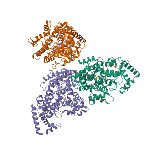
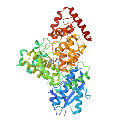
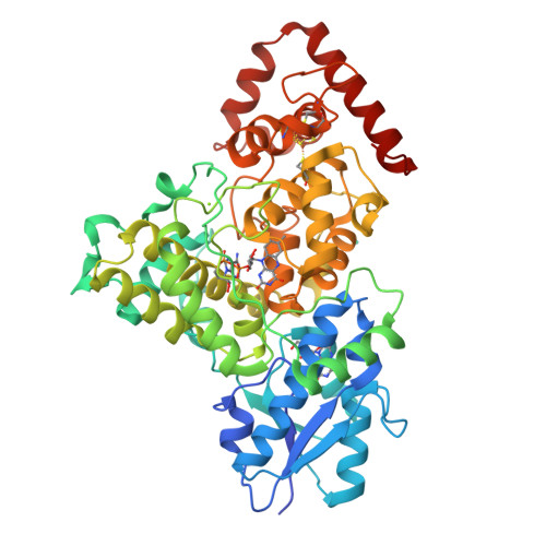
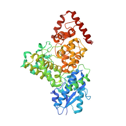
    Cryptochromes and photolyases are structurally related but have different biological functions in signalling and DNA repair. Proteobacteria and cyanobacteria harbour a new class of cryptochromes, called CryPro. We have solved the 2.7 Å structure of one of its members, cryptochrome B from Rhodobacter sphaeroides, which is a regulator of photosynthesis gene expression. The structure reveals that, in addition to the photolyase-like fold, CryB contains two cofactors only conserved in the CryPro subfamily: 6,7-dimethyl-8-ribityl-lumazine in the antenna-binding domain and a [4Fe-4S] cluster within the catalytic domain. The latter closely resembles the iron-sulphur cluster harbouring the large primase subunit PriL, indicating that PriL is evolutionarily related to the CryPro class of cryptochromes.


  • Organizational Affiliation

    Department of Chemistry-Institute of Biochemistry, Philipps-University, Marburg, Germany.


Macromolecules
Find similar proteins by:  (by identity cutoff)  |  3D Structure
Entity ID: 1
MoleculeChains Sequence LengthOrganismDetailsImage
CRYPTOCHROME B
A, B, C
522Cereibacter sphaeroides 2.4.1Mutation(s): 0 
UniProt
Find proteins for Q3IXP1 (Cereibacter sphaeroides (strain ATCC 17023 / DSM 158 / JCM 6121 / CCUG 31486 / LMG 2827 / NBRC 12203 / NCIMB 8253 / ATH 2.4.1.))
Explore Q3IXP1 
Go to UniProtKB:  Q3IXP1
Entity Groups  
Sequence Clusters30% Identity50% Identity70% Identity90% Identity95% Identity100% Identity
UniProt GroupQ3IXP1
Sequence Annotations
Expand
  • Reference Sequence
Small Molecules
Ligands 5 Unique
IDChains Name / Formula / InChI Key2D Diagram3D Interactions
FAD
Query on FAD

Download Ideal Coordinates CCD File 
D [auth A],
I [auth B],
N [auth C]
FLAVIN-ADENINE DINUCLEOTIDE
C27 H33 N9 O15 P2
VWWQXMAJTJZDQX-UYBVJOGSSA-N
SF4
Query on SF4

Download Ideal Coordinates CCD File 
E [auth A],
J [auth B],
O [auth C]
IRON/SULFUR CLUSTER
Fe4 S4
LJBDFODJNLIPKO-UHFFFAOYSA-N
DLZ
Query on DLZ

Download Ideal Coordinates CCD File 
F [auth A],
K [auth B],
P [auth C]
1-deoxy-1-(6,7-dimethyl-2,4-dioxo-3,4-dihydropteridin-8(2H)-yl)-D-ribitol
C13 H18 N4 O6
SXDXRJZUAJBNFL-XKSSXDPKSA-N
GD
Query on GD

Download Ideal Coordinates CCD File 
H [auth A],
M [auth B],
R [auth C]
GADOLINIUM ATOM
Gd
UIWYJDYFSGRHKR-UHFFFAOYSA-N
MG
Query on MG

Download Ideal Coordinates CCD File 
G [auth A],
L [auth B],
Q [auth C]
MAGNESIUM ION
Mg
JLVVSXFLKOJNIY-UHFFFAOYSA-N
Experimental Data & Validation

Experimental Data

  • Method: X-RAY DIFFRACTION
  • Resolution: 2.70 Å
  • R-Value Free:  0.228 (Depositor), 0.220 (DCC) 
  • R-Value Work:  0.198 (Depositor), 0.200 (DCC) 
  • R-Value Observed: 0.199 (Depositor) 
Space Group: P 65 2 2
Unit Cell:
Length ( Å )Angle ( ˚ )
a = 137.84α = 90
b = 137.84β = 90
c = 521.94γ = 120
Software Package:
Software NamePurpose
REFMACrefinement
XDSdata reduction
XSCALEdata scaling
PHENIXphasing

Structure Validation

View Full Validation Report


Ligand Structure Quality Assessment 

Created with Raphaël 2.3.0Worse 01 BetterLigand structure goodness of fit to experimental dataBest fitted FADClick on this verticalbar to view detailsBest fitted DLZClick on this verticalbar to view details

Entry History 

Deposition Data

Revision History  (Full details and data files)

  • Version 1.0: 2012-02-15
    Type: Initial release
  • Version 1.1: 2012-03-28
    Changes: Other
  • Version 1.2: 2014-08-13
    Changes: Refinement description
  • Version 1.3: 2024-05-08
    Changes: Data collection, Database references, Derived calculations, Other, Refinement description