4DBC | pdb_00004dbc

Substrate Activation in Aspartate Aminotransferase


Experimental Data Snapshot

  • Method: X-RAY DIFFRACTION
  • Resolution: 1.50 Å
  • R-Value Free: 
    0.194 (Depositor), 0.190 (DCC) 
  • R-Value Work: 
    0.169 (Depositor), 0.160 (DCC) 
  • R-Value Observed: 
    0.170 (Depositor) 

wwPDB Validation   3D Report Full Report


Ligand Structure Quality Assessment 

Created with Raphaël 2.3.0Worse 01 BetterLigand structure goodness of fit to experimental dataBest fitted 3QPClick on this verticalbar to view details

This is version 1.2 of the entry. See complete history


Literature

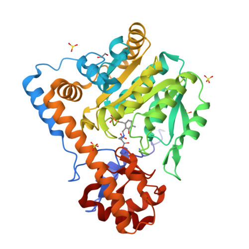
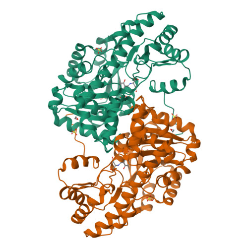
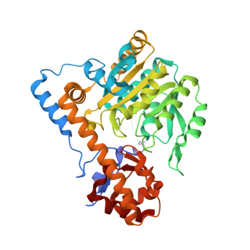
Ground-state electronic destabilization via hyperconjugation in aspartate aminotransferase.

Griswold, W.R.Castro, J.N.Fisher, A.J.Toney, M.D.

(2012) J Am Chem Soc 134: 8436-8438

  • DOI: https://doi.org/10.1021/ja302809e
  • Primary Citation of Related Structures:  
    4DBC

  • PubMed Abstract: 

    Binding isotope effects for l-aspartate reacting with the inactive K258A mutant of PLP-dependent aspartate aminotransferase to give a stable external aldimine intermediate are reported. They provide direct evidence for electronic ground-state destabilization via hyperconjugation. The smaller equilibrium isotope effect with deazaPLP-reconstituted K258A indicates that the pyridine nitrogen plays an important role in labilizing the Cα-H bond.


  • Organizational Affiliation

    Department of Chemistry, University of California - Davis, 95616, United States.


Macromolecules
Find similar proteins by:  (by identity cutoff)  |  3D Structure
Entity ID: 1
MoleculeChains Sequence LengthOrganismDetailsImage
Aspartate aminotransferase396Escherichia coli K-12Mutation(s): 1 
EC: 2.6.1.1
UniProt
Find proteins for P00509 (Escherichia coli (strain K12))
Explore P00509 
Go to UniProtKB:  P00509
Entity Groups  
Sequence Clusters30% Identity50% Identity70% Identity90% Identity95% Identity100% Identity
UniProt GroupP00509
Sequence Annotations
Expand
  • Reference Sequence
Small Molecules
Ligands 3 Unique
IDChains Name / Formula / InChI Key2D Diagram3D Interactions
3QP
Query on 3QP

Download Ideal Coordinates CCD File 
G [auth A](E)-N-{2-hydroxy-3-methyl-6-[(phosphonooxy)methyl]benzylidene}-L-aspartic acid
C13 H16 N O9 P
CMDDKMGWJXISQE-ZFGNZVLRSA-N
SO4
Query on SO4

Download Ideal Coordinates CCD File 
B [auth A],
C [auth A],
D [auth A],
E [auth A]
SULFATE ION
O4 S
QAOWNCQODCNURD-UHFFFAOYSA-L
EDO
Query on EDO

Download Ideal Coordinates CCD File 
F [auth A]1,2-ETHANEDIOL
C2 H6 O2
LYCAIKOWRPUZTN-UHFFFAOYSA-N
Experimental Data & Validation

Experimental Data

  • Method: X-RAY DIFFRACTION
  • Resolution: 1.50 Å
  • R-Value Free:  0.194 (Depositor), 0.190 (DCC) 
  • R-Value Work:  0.169 (Depositor), 0.160 (DCC) 
  • R-Value Observed: 0.170 (Depositor) 
Space Group: C 2 2 21
Unit Cell:
Length ( Å )Angle ( ˚ )
a = 84.29α = 90
b = 155.25β = 90
c = 77.96γ = 90
Software Package:
Software NamePurpose
MOLREPphasing
PHENIXrefinement
DENZOdata reduction
SCALAdata scaling

Structure Validation

View Full Validation Report


Ligand Structure Quality Assessment 

Created with Raphaël 2.3.0Worse 01 BetterLigand structure goodness of fit to experimental dataBest fitted 3QPClick on this verticalbar to view details

Entry History 

Deposition Data

Revision History  (Full details and data files)

  • Version 1.0: 2012-12-05
    Type: Initial release
  • Version 1.1: 2018-08-29
    Changes: Data collection, Source and taxonomy, Structure summary
  • Version 1.2: 2024-02-28
    Changes: Data collection, Database references, Derived calculations