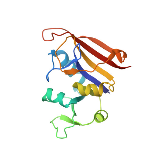
Stereo-selectively Induced Cofactor Switching Provides Insight into Cofactor Site Plasticity as a Possible Mechanism of Antifolate Resistance
Keshipeddy, S., Reeve, S.M., Anderson, A.C., Wright, D.L.To be published.
Experimental Data Snapshot
Starting Model: experimental
View more details
Entity ID: 1 | |||||
|---|---|---|---|---|---|
| Molecule | Chains | Sequence Length | Organism | Details | Image |
| Dihydrofolate reductase | A [auth X] | 160 | Staphylococcus aureus | Mutation(s): 0 Gene Names: folA EC: 1.5.1.3 |  |
UniProt | |||||
Find proteins for P0A017 (Staphylococcus aureus) Explore P0A017 Go to UniProtKB: P0A017 | |||||
Entity Groups | |||||
| Sequence Clusters | 30% Identity50% Identity70% Identity90% Identity95% Identity100% Identity | ||||
| UniProt Group | P0A017 | ||||
Sequence AnnotationsExpand | |||||
| |||||
| Ligands 3 Unique | |||||
|---|---|---|---|---|---|
| ID | Chains | Name / Formula / InChI Key | 2D Diagram | 3D Interactions | |
| NDP Query on NDP | D [auth X] | NADPH DIHYDRO-NICOTINAMIDE-ADENINE-DINUCLEOTIDE PHOSPHATE C21 H30 N7 O17 P3 ACFIXJIJDZMPPO-NNYOXOHSSA-N |  | ||
| 06U Query on 06U | B [auth X] | 6-ethyl-5-{(3R)-3-[3-methoxy-5-(pyridin-4-yl)phenyl]but-1-yn-1-yl}pyrimidine-2,4-diamine C22 H23 N5 O KEPLBUUTAQCZOE-AWEZNQCLSA-N |  | ||
| ACT Query on ACT | C [auth X] | ACETATE ION C2 H3 O2 QTBSBXVTEAMEQO-UHFFFAOYSA-M |  | ||
| Length ( Å ) | Angle ( ˚ ) |
|---|---|
| a = 78.902 | α = 90 |
| b = 78.902 | β = 90 |
| c = 108.118 | γ = 120 |
| Software Name | Purpose |
|---|---|
| PHENIX | refinement |
| HKL-2000 | data reduction |
| HKL-2000 | data scaling |
| PHENIX | phasing |
| Funding Organization | Location | Grant Number |
|---|---|---|
| National Institutes of Health/National Institute Of Allergy and Infectious Diseases (NIH/NIAID) | United States | AI111957 |