RCSB PDB - 5F1B: Structural basis of Ebola virus entry: viral glycoprotein bound to its endosomal receptor Niemann-Pick C1

 5F1B

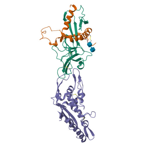
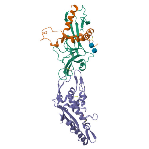
Structural basis of Ebola virus entry: viral glycoprotein bound to its endosomal receptor Niemann-Pick C1


Experimental Data Snapshot

  • Method: X-RAY DIFFRACTION
  • Resolution: 2.30 Å
  • R-Value Free: 0.230 
  • R-Value Work: 0.181 
  • R-Value Observed: 0.184 

Starting Model: experimental
View more details

wwPDB Validation   3D Report Full Report


This is version 2.2 of the entry. See complete history


Literature

Ebola Viral Glycoprotein Bound to Its Endosomal Receptor Niemann-Pick C1.

Wang, H.Shi, Y.Song, J.Qi, J.Lu, G.Yan, J.Gao, G.F.

(2016) Cell 164: 258-268

  • DOI: https://doi.org/10.1016/j.cell.2015.12.044
  • Primary Citation of Related Structures:  
    5F18, 5F1B

  • PubMed Abstract: 

    Filoviruses, including Ebola and Marburg, cause fatal hemorrhagic fever in humans and primates. Understanding how these viruses enter host cells could help to develop effective therapeutics. An endosomal protein, Niemann-Pick C1 (NPC1), has been identified as a necessary entry receptor for this process, and priming of the viral glycoprotein (GP) to a fusion-competent state is a prerequisite for NPC1 binding. Here, we have determined the crystal structure of the primed GP (GPcl) of Ebola virus bound to domain C of NPC1 (NPC1-C) at a resolution of 2.3 Å. NPC1-C utilizes two protruding loops to engage a hydrophobic cavity on head of GPcl. Upon enzymatic cleavage and NPC1-C binding, conformational change in the GPcl further affects the state of the internal fusion loop, triggering membrane fusion. Our data therefore provide structural insights into filovirus entry in the late endosome and the molecular basis for design of therapeutic inhibitors of viral entry.


  • Organizational Affiliation

    CAS Key Laboratory of Pathogenic Microbiology and Immunology, Institute of Microbiology, Chinese Academy of Sciences, Beijing 100101, China; University of Chinese Academy of Sciences, Beijing 100049, China.


Macromolecules
Find similar proteins by:  (by identity cutoff)  |  3D Structure
Entity ID: 1
MoleculeChains Sequence LengthOrganismDetailsImage
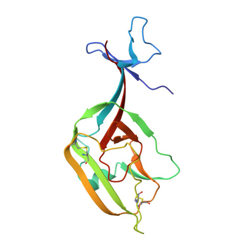
GP1158Ebola virus - Zaire (1995)Mutation(s): 1 
Gene Names: GP
Membrane Entity: Yes 
UniProt
Find proteins for P87666 (Zaire ebolavirus (strain Kikwit-95))
Explore P87666 
Go to UniProtKB:  P87666
Entity Groups  
Sequence Clusters30% Identity50% Identity70% Identity90% Identity95% Identity100% Identity
UniProt GroupP87666
Sequence Annotations
Expand
  • Reference Sequence
Find similar proteins by:  (by identity cutoff)  |  3D Structure
Entity ID: 2
MoleculeChains Sequence LengthOrganismDetailsImage
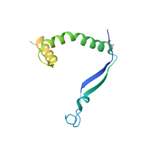
GP2130Ebola virus - Zaire (1995)Mutation(s): 0 
Gene Names: GP
Membrane Entity: Yes 
UniProt
Find proteins for P87666 (Zaire ebolavirus (strain Kikwit-95))
Explore P87666 
Go to UniProtKB:  P87666
Entity Groups  
Sequence Clusters30% Identity50% Identity70% Identity90% Identity95% Identity100% Identity
UniProt GroupP87666
Glycosylation
Glycosylation Sites: 1
Sequence Annotations
Expand
  • Reference Sequence
Find similar proteins by:  (by identity cutoff)  |  3D Structure
Entity ID: 3
MoleculeChains Sequence LengthOrganismDetailsImage
Niemann-Pick C1 protein256Homo sapiensMutation(s): 0 
Gene Names: NPC1
Membrane Entity: Yes 
UniProt & NIH Common Fund Data Resources
Find proteins for O15118 (Homo sapiens)
Explore O15118 
Go to UniProtKB:  O15118
PHAROS:  O15118
GTEx:  ENSG00000141458 
Entity Groups  
Sequence Clusters30% Identity50% Identity70% Identity90% Identity95% Identity100% Identity
UniProt GroupO15118
Sequence Annotations
Expand
  • Reference Sequence
Oligosaccharides

Help

Entity ID: 4
MoleculeChains Length2D Diagram Glycosylation3D Interactions
2-acetamido-2-deoxy-beta-D-glucopyranose-(1-4)-2-acetamido-2-deoxy-beta-D-glucopyranose
D
2N-Glycosylation
Glycosylation Resources
GlyTouCan:  G42666HT
GlyCosmos:  G42666HT
GlyGen:  G42666HT
Experimental Data & Validation

Experimental Data

  • Method: X-RAY DIFFRACTION
  • Resolution: 2.30 Å
  • R-Value Free: 0.230 
  • R-Value Work: 0.181 
  • R-Value Observed: 0.184 
  • Space Group: P 63
Unit Cell:
Length ( Å )Angle ( ˚ )
a = 107.261α = 90
b = 107.261β = 90
c = 93.777γ = 120
Software Package:
Software NamePurpose
PHENIXrefinement
HKL-3000data reduction
HKL-3000data scaling
PHASERphasing

Structure Validation

View Full Validation Report


Entry History & Funding Information

Deposition Data


Funding OrganizationLocationGrant Number
China Ministry of Science and Technology National 973 ProjectChina2013CB531502 and 2014CB542503

Revision History  (Full details and data files)

  • Version 1.0: 2016-01-20
    Type: Initial release
  • Version 1.1: 2016-01-27
    Changes: Database references
  • Version 2.0: 2020-07-29
    Type: Remediation
    Reason: Carbohydrate remediation
    Changes: Advisory, Atomic model, Data collection, Derived calculations, Structure summary
  • Version 2.1: 2023-11-08
    Changes: Data collection, Database references, Refinement description, Structure summary
  • Version 2.2: 2024-10-09
    Changes: Structure summary