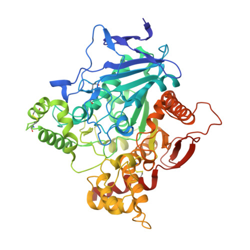
5HFA

Crystal structure of human acetylcholinesterase in complex with paraoxon and 2-PAM


Experimental Data Snapshot

  • Method: X-RAY DIFFRACTION
  • Resolution: 2.20 Å
  • R-Value Free: 0.224 
  • R-Value Work: 0.181 
  • R-Value Observed: 0.183 

Starting Model: experimental
View more details

wwPDB Validation   3D Report Full Report


Ligand Structure Quality Assessment 


This is version 2.2 of the entry. See complete history


Literature

Structures of paraoxon-inhibited human acetylcholinesterase reveal perturbations of the acyl loop and the dimer interface.

Franklin, M.C.Rudolph, M.J.Ginter, C.Cassidy, M.S.Cheung, J.

(2016) Proteins 84: 1246-1256

  • DOI: https://doi.org/10.1002/prot.25073
  • Primary Citation of Related Structures:  
    5HF5, 5HF6, 5HF8, 5HF9, 5HFA

  • PubMed Abstract: 

    Irreversible inhibition of the essential nervous system enzyme acetylcholinesterase by organophosphate nerve agents and pesticides may quickly lead to death. Oxime reactivators currently used as antidotes are generally less effective against pesticide exposure than nerve agent exposure, and pesticide exposure constitutes the majority of cases of organophosphate poisoning in the world. The current lack of published structural data specific to human acetylcholinesterase organophosphate-inhibited and oxime-bound states hinders development of effective medical treatments. We have solved structures of human acetylcholinesterase in different states in complex with the organophosphate insecticide, paraoxon, and oximes. Reaction with paraoxon results in a highly perturbed acyl loop that causes a narrowing of the gorge in the peripheral site that may impede entry of reactivators. This appears characteristic of acetylcholinesterase inhibition by organophosphate insecticides but not nerve agents. Additional changes seen at the dimer interface are novel and provide further examples of the disruptive effect of paraoxon. Ternary structures of paraoxon-inhibited human acetylcholinesterase in complex with the oximes HI6 and 2-PAM reveals relatively poor positioning for reactivation. This study provides a structural foundation for improved reactivator design for the treatment of organophosphate intoxication. Proteins 2016; 84:1246-1256. © 2016 Wiley Periodicals, Inc.


  • Organizational Affiliation

    Special Projects Group, New York Structural Biology Center, New York, New York, 10027.


Macromolecules
Find similar proteins by:  (by identity cutoff)  |  3D Structure
Entity ID: 1
MoleculeChains Sequence LengthOrganismDetailsImage
Acetylcholinesterase
A, B
542Homo sapiensMutation(s): 0 
Gene Names: ACHE
EC: 3.1.1.7
UniProt & NIH Common Fund Data Resources
Find proteins for P22303 (Homo sapiens)
Explore P22303 
Go to UniProtKB:  P22303
PHAROS:  P22303
GTEx:  ENSG00000087085 
Entity Groups  
Sequence Clusters30% Identity50% Identity70% Identity90% Identity95% Identity100% Identity
UniProt GroupP22303
Glycosylation
Glycosylation Sites: 2Go to GlyGen: P22303-1
Sequence Annotations
Expand
  • Reference Sequence
Oligosaccharides

Help

Entity ID: 2
MoleculeChains Length2D Diagram Glycosylation3D Interactions
2-acetamido-2-deoxy-beta-D-glucopyranose-(1-4)-[alpha-L-fucopyranose-(1-6)]2-acetamido-2-deoxy-beta-D-glucopyranose
C, D
3N-Glycosylation
Glycosylation Resources
GlyTouCan:  G21290RB
GlyCosmos:  G21290RB
GlyGen:  G21290RB
Small Molecules
Ligands 5 Unique
IDChains Name / Formula / InChI Key2D Diagram3D Interactions
NAG
Query on NAG

Download Ideal Coordinates CCD File 
J [auth B]2-acetamido-2-deoxy-beta-D-glucopyranose
C8 H15 N O6
OVRNDRQMDRJTHS-FMDGEEDCSA-N
FP1
Query on FP1

Download Ideal Coordinates CCD File 
H [auth A],
I [auth A],
O [auth B]
N-hydroxy-1-(1-methylpyridin-2(1H)-ylidene)methanamine
C7 H10 N2 O
YDWKOUCMHAMECR-SREVYHEPSA-N
DEP
Query on DEP

Download Ideal Coordinates CCD File 
E [auth A],
K [auth B]
DIETHYL PHOSPHONATE
C4 H11 O3 P
MJUJXFBTEFXVKU-UHFFFAOYSA-N
EDO
Query on EDO

Download Ideal Coordinates CCD File 
F [auth A],
G [auth A],
L [auth B],
M [auth B]
1,2-ETHANEDIOL
C2 H6 O2
LYCAIKOWRPUZTN-UHFFFAOYSA-N
NO3
Query on NO3

Download Ideal Coordinates CCD File 
N [auth B]NITRATE ION
N O3
NHNBFGGVMKEFGY-UHFFFAOYSA-N
Experimental Data & Validation

Experimental Data

  • Method: X-RAY DIFFRACTION
  • Resolution: 2.20 Å
  • R-Value Free: 0.224 
  • R-Value Work: 0.181 
  • R-Value Observed: 0.183 
  • Space Group: P 31 2 1
Unit Cell:
Length ( Å )Angle ( ˚ )
a = 104.803α = 90
b = 104.803β = 90
c = 323.462γ = 120
Software Package:
Software NamePurpose
HKL-2000data collection
SCALEPACKdata scaling
PHENIXrefinement
PDB_EXTRACTdata extraction
HKL-2000data scaling

Structure Validation

View Full Validation Report


Ligand Structure Quality Assessment 


Entry History 

Deposition Data

Revision History  (Full details and data files)

  • Version 1.0: 2016-06-22
    Type: Initial release
  • Version 1.1: 2016-08-24
    Changes: Database references
  • Version 1.2: 2017-11-22
    Changes: Database references, Derived calculations, Refinement description
  • Version 2.0: 2020-07-29
    Type: Remediation
    Reason: Carbohydrate remediation
    Changes: Atomic model, Data collection, Derived calculations, Structure summary
  • Version 2.1: 2023-09-27
    Changes: Data collection, Database references, Derived calculations, Refinement description, Structure summary
  • Version 2.2: 2024-10-30
    Changes: Structure summary