5ZK4 | pdb_00005zk4

The structure of DSZS acyltransferase with carrier protein


Experimental Data Snapshot

  • Method: X-RAY DIFFRACTION
  • Resolution: 2.03 Å
  • R-Value Free: 
    0.222 (Depositor), 0.230 (DCC) 
  • R-Value Work: 
    0.183 (Depositor), 0.190 (DCC) 
  • R-Value Observed: 
    0.185 (Depositor) 

Starting Model: experimental
View more details

wwPDB Validation   3D Report Full Report


Ligand Structure Quality Assessment 

Created with Raphaël 2.3.0Worse 01 BetterLigand structure goodness of fit to experimental dataBest fitted 9EFClick on this verticalbar to view details

This is version 1.4 of the entry. See complete history


Literature

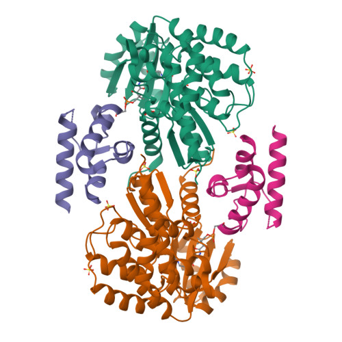
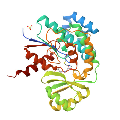
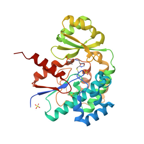
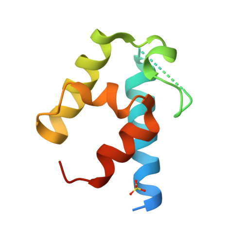
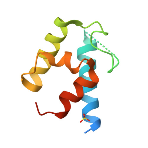
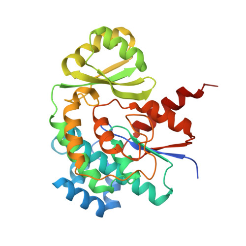
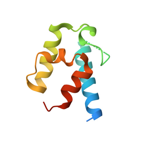
Structural basis of protein-protein interactions between a trans-acting acyltransferase and acyl carrier protein in polyketide disorazole biosynthesis

Miyanaga, A.Ouchi, R.Ishikawa, F.Goto, E.Tanabe, G.Kudo, F.Eguchi, T.

(2018) J Am Chem Soc 140: 7970-7978

  • DOI: https://doi.org/10.1021/jacs.8b04162
  • Primary Citation of Related Structures:  
    5ZK4

  • PubMed Abstract: 

    Acyltransferases (ATs) are responsible for the selection and incorporation of acyl building blocks in the biosynthesis of various polyketide natural products. The trans-AT modular polyketide synthases have a discrete trans-acting AT for the loading of an acyl unit onto the acyl carrier protein (ACP) located within each module. Despite the importance of protein-protein interactions between ATs and ACPs in trans-AT assembly lines, the dynamic actions of ACPs and trans-acting ATs remain largely uncharacterized because of the inherently transient nature of ACP-enzyme interactions. Herein, we report the crystal structure of the AT-ACP complex of disorazole trans-AT polyketide synthase. We used a bromoacetamide pantetheine cross-linking probe in combination with a Cys mutation to trap the transient AT-ACP complex, allowing the determination of the crystal structure of the disorazole AT-ACP complex at 2.03 Å resolution. On the basis of the cross-linked AT-ACP complex structure, ACP residues recognized by trans-acting AT were identified and validated by mutational studies, which demonstrated that the disorazole AT recognizes the loop 1 and helix III' residues of disorazole ACP. The disorazole AT-ACP complex structure presents a foundation for defining the dynamic processes associated with trans-acting ATs and provides detailed mechanistic insights into their ability to recognize ACPs.


  • Organizational Affiliation

    Department of Chemistry , Tokyo Institute of Technology , 2-12-1, O-okayama , Meguro-ku, Tokyo 152-8551 , Japan.


Macromolecules
Find similar proteins by:  (by identity cutoff)  |  3D Structure
Entity ID: 1
MoleculeChains Sequence LengthOrganismDetailsImage
DisD protein
A, B
302Sorangium cellulosumMutation(s): 1 
Gene Names: dszDdisD
EC: 2.3.1.39
UniProt
Find proteins for Q4U443 (Sorangium cellulosum)
Explore Q4U443 
Go to UniProtKB:  Q4U443
Entity Groups  
Sequence Clusters30% Identity50% Identity70% Identity90% Identity95% Identity100% Identity
UniProt GroupQ4U443
Sequence Annotations
Expand
  • Reference Sequence
Find similar proteins by:  (by identity cutoff)  |  3D Structure
Entity ID: 2
MoleculeChains Sequence LengthOrganismDetailsImage
DisA protein
C, D
94Sorangium cellulosumMutation(s): 0 
Gene Names: dszAdisA
UniProt
Find proteins for Q4U447 (Sorangium cellulosum)
Explore Q4U447 
Go to UniProtKB:  Q4U447
Entity Groups  
Sequence Clusters30% Identity50% Identity70% Identity90% Identity95% Identity100% Identity
UniProt GroupQ4U447
Sequence Annotations
Expand
  • Reference Sequence
Small Molecules
Ligands 2 Unique
IDChains Name / Formula / InChI Key2D Diagram3D Interactions
9EF
Query on 9EF

Download Ideal Coordinates CCD File 
I [auth A],
N [auth B]
N-[2-(acetylamino)ethyl]-N~3~-[(2R)-2-hydroxy-3,3-dimethyl-4-(phosphonooxy)butanoyl]-beta-alaninamide
C13 H26 N3 O8 P
GTANJSAWPNEGPZ-NSHDSACASA-N
SO4
Query on SO4

Download Ideal Coordinates CCD File 
E [auth A]
F [auth A]
G [auth A]
H [auth A]
J [auth B]
SULFATE ION
O4 S
QAOWNCQODCNURD-UHFFFAOYSA-L
Experimental Data & Validation

Experimental Data

  • Method: X-RAY DIFFRACTION
  • Resolution: 2.03 Å
  • R-Value Free:  0.222 (Depositor), 0.230 (DCC) 
  • R-Value Work:  0.183 (Depositor), 0.190 (DCC) 
  • R-Value Observed: 0.185 (Depositor) 
Space Group: P 21 21 21
Unit Cell:
Length ( Å )Angle ( ˚ )
a = 90.324α = 90
b = 96.629β = 90
c = 99.056γ = 90
Software Package:
Software NamePurpose
REFMACrefinement
XDSdata reduction
SCALAdata scaling
MOLREPphasing
ARP/wARPmodel building
Cootmodel building

Structure Validation

View Full Validation Report


Ligand Structure Quality Assessment 

Created with Raphaël 2.3.0Worse 01 BetterLigand structure goodness of fit to experimental dataBest fitted 9EFClick on this verticalbar to view details

Entry History & Funding Information

Deposition Data


Funding OrganizationLocationGrant Number
Japan Society for the Promotion of ScienceJapan17K07747

Revision History  (Full details and data files)

  • Version 1.0: 2018-06-13
    Type: Initial release
  • Version 1.1: 2018-06-20
    Changes: Data collection, Database references
  • Version 1.2: 2018-07-04
    Changes: Data collection, Database references
  • Version 1.3: 2023-11-22
    Changes: Data collection, Database references, Derived calculations, Refinement description
  • Version 1.4: 2024-11-13
    Changes: Structure summary