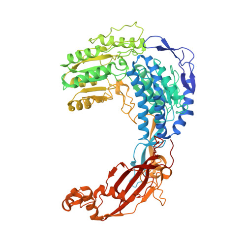
6JQM

Structure of PaaZ with NADPH


Experimental Data Snapshot

  • Method: ELECTRON MICROSCOPY
  • Resolution: 3.30 Å
  • Aggregation State: PARTICLE 
  • Reconstruction Method: SINGLE PARTICLE 

wwPDB Validation   3D Report Full Report


This is version 1.3 of the entry. See complete history


Literature

Molecular basis for metabolite channeling in a ring opening enzyme of the phenylacetate degradation pathway.

Sathyanarayanan, N.Cannone, G.Gakhar, L.Katagihallimath, N.Sowdhamini, R.Ramaswamy, S.Vinothkumar, K.R.

(2019) Nat Commun 10: 4127-4127

  • DOI: https://doi.org/10.1038/s41467-019-11931-1
  • Primary Citation of Related Structures:  
    6JQL, 6JQM, 6JQN, 6JQO

  • PubMed Abstract: 

    Substrate channeling is a mechanism for the internal transfer of hydrophobic, unstable or toxic intermediates from the active site of one enzyme to another. Such transfer has previously been described to be mediated by a hydrophobic tunnel, the use of electrostatic highways or pivoting and by conformational changes. The enzyme PaaZ is used by many bacteria to degrade environmental pollutants. PaaZ is a bifunctional enzyme that catalyzes the ring opening of oxepin-CoA and converts it to 3-oxo-5,6-dehydrosuberyl-CoA. Here we report the structures of PaaZ determined by electron cryomicroscopy with and without bound ligands. The structures reveal that three domain-swapped dimers of the enzyme form a trilobed structure. A combination of small-angle X-ray scattering (SAXS), computational studies, mutagenesis and microbial growth experiments suggests that the key intermediate is transferred from one active site to the other by a mechanism of electrostatic pivoting of the CoA moiety, mediated by a set of conserved positively charged residues.


  • Organizational Affiliation

    Institute for Stem Cell Science and Regenerative Medicine, GKVK Campus, Bellary Road, Bangalore, India.


Macromolecules
Find similar proteins by:  (by identity cutoff)  |  3D Structure
Entity ID: 1
MoleculeChains Sequence LengthOrganismDetailsImage
Bifunctional protein PaaZ
A, B, C, D, E
A, B, C, D, E, F
688Escherichia coli K-12Mutation(s): 0 
Gene Names: paaZ
EC: 3.3.2.12 (PDB Primary Data), 1.2.1.91 (UniProt)
UniProt
Find proteins for P77455 (Escherichia coli (strain K12))
Explore P77455 
Go to UniProtKB:  P77455
Entity Groups  
Sequence Clusters30% Identity50% Identity70% Identity90% Identity95% Identity100% Identity
UniProt GroupP77455
Sequence Annotations
Expand
  • Reference Sequence
Experimental Data & Validation

Experimental Data

  • Method: ELECTRON MICROSCOPY
  • Resolution: 3.30 Å
  • Aggregation State: PARTICLE 
  • Reconstruction Method: SINGLE PARTICLE 
EM Software:
TaskSoftware PackageVersion
MODEL REFINEMENTPHENIX1.13
RECONSTRUCTIONRELION2.0

Structure Validation

View Full Validation Report


Entry History & Funding Information

Deposition Data


Funding OrganizationLocationGrant Number
Department of Biotechnology (India)IndiaDBT/PR12422/MED/31/287/2014
Council of Scientific & Industrial ResearchIndiaCSIR/37/1606/13/EMR-II
Department of Biotechnology (India)IndiaBT/PR5081/INF/22/156/2012
Medical Research Council (United Kingdom)United KingdomU105184322

Revision History  (Full details and data files)

  • Version 1.0: 2019-09-11
    Type: Initial release
  • Version 1.1: 2019-09-18
    Changes: Data collection, Database references
  • Version 1.2: 2019-09-25
    Changes: Data collection, Database references
  • Version 1.3: 2024-03-27
    Changes: Data collection, Database references