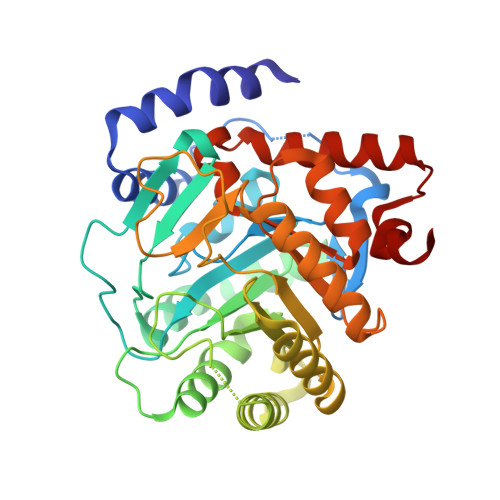
6QU7

Crystal structure of human DHODH in complex with BAY 2402234


Experimental Data Snapshot

  • Method: X-RAY DIFFRACTION
  • Resolution: 1.52 Å
  • R-Value Free: 0.173 
  • R-Value Work: 0.152 
  • R-Value Observed: 0.152 

wwPDB Validation   3D Report Full Report


Ligand Structure Quality Assessment 


This is version 1.2 of the entry. See complete history


Literature

The novel dihydroorotate dehydrogenase (DHODH) inhibitor BAY 2402234 triggers differentiation and is effective in the treatment of myeloid malignancies.

Christian, S.Merz, C.Evans, L.Gradl, S.Seidel, H.Friberg, A.Eheim, A.Lejeune, P.Brzezinka, K.Zimmermann, K.Ferrara, S.Meyer, H.Lesche, R.Stoeckigt, D.Bauser, M.Haegebarth, A.Sykes, D.B.Scadden, D.T.Losman, J.A.Janzer, A.

(2019) Leukemia 33: 2403-2415

  • DOI: https://doi.org/10.1038/s41375-019-0461-5
  • Primary Citation of Related Structures:  
    6QU7

  • PubMed Abstract: 

    Acute myeloid leukemia (AML) is a devastating disease, with the majority of patients dying within a year of diagnosis. For patients with relapsed/refractory AML, the prognosis is particularly poor with currently available treatments. Although genetically heterogeneous, AML subtypes share a common differentiation arrest at hematopoietic progenitor stages. Overcoming this differentiation arrest has the potential to improve the long-term survival of patients, as is the case in acute promyelocytic leukemia (APL), which is characterized by a chromosomal translocation involving the retinoic acid receptor alpha gene. Treatment of APL with all-trans retinoic acid (ATRA) induces terminal differentiation and apoptosis of leukemic promyelocytes, resulting in cure rates of over 80%. Unfortunately, similarly efficacious differentiation therapies have, to date, been lacking outside of APL. Inhibition of dihydroorotate dehydrogenase (DHODH), a key enzyme in the de novo pyrimidine synthesis pathway, was recently reported to induce differentiation of diverse AML subtypes. In this report we describe the discovery and characterization of BAY 2402234 - a novel, potent, selective and orally bioavailable DHODH inhibitor that shows monotherapy efficacy and differentiation induction across multiple AML subtypes. Herein, we present the preclinical data that led to initiation of a phase I evaluation of this inhibitor in myeloid malignancies.


  • Organizational Affiliation

    Bayer AG, Muellerstrasse 178, 13353, Berlin, Germany.


Macromolecules
Find similar proteins by:  (by identity cutoff)  |  3D Structure
Entity ID: 1
MoleculeChains Sequence LengthOrganismDetailsImage
Dihydroorotate dehydrogenase (quinone), mitochondrial366Homo sapiensMutation(s): 0 
Gene Names: DHODH
EC: 1.3.5.2
UniProt & NIH Common Fund Data Resources
Find proteins for Q02127 (Homo sapiens)
Explore Q02127 
Go to UniProtKB:  Q02127
PHAROS:  Q02127
GTEx:  ENSG00000102967 
Entity Groups  
Sequence Clusters30% Identity50% Identity70% Identity90% Identity95% Identity100% Identity
UniProt GroupQ02127
Sequence Annotations
Expand
  • Reference Sequence
Small Molecules
Ligands 5 Unique
IDChains Name / Formula / InChI Key2D Diagram3D Interactions
JJE
Query on JJE

Download Ideal Coordinates CCD File 
D [auth A]~{N}-(2-chloranyl-6-fluoranyl-phenyl)-4-[4-ethyl-3-(hydroxymethyl)-5-oxidanylidene-1,2,4-triazol-1-yl]-5-fluoranyl-2-[(2~{S})-1,1,1-tris(fluoranyl)propan-2-yl]oxy-benzamide
C21 H18 Cl F5 N4 O4
KNVJMHHAXCPZHF-JTQLQIEISA-N
FMN
Query on FMN

Download Ideal Coordinates CCD File 
C [auth A]FLAVIN MONONUCLEOTIDE
C17 H21 N4 O9 P
FVTCRASFADXXNN-SCRDCRAPSA-N
ORO
Query on ORO

Download Ideal Coordinates CCD File 
B [auth A]OROTIC ACID
C5 H4 N2 O4
PXQPEWDEAKTCGB-UHFFFAOYSA-N
SO4
Query on SO4

Download Ideal Coordinates CCD File 
E [auth A],
F [auth A]
SULFATE ION
O4 S
QAOWNCQODCNURD-UHFFFAOYSA-L
ACT
Query on ACT

Download Ideal Coordinates CCD File 
G [auth A],
H [auth A],
I [auth A],
J [auth A]
ACETATE ION
C2 H3 O2
QTBSBXVTEAMEQO-UHFFFAOYSA-M
Binding Affinity Annotations 
IDSourceBinding Affinity
JJE BindingDB:  6QU7 IC50: min: 1.2, max: 6 (nM) from 4 assay(s)
Experimental Data & Validation

Experimental Data

  • Method: X-RAY DIFFRACTION
  • Resolution: 1.52 Å
  • R-Value Free: 0.173 
  • R-Value Work: 0.152 
  • R-Value Observed: 0.152 
  • Space Group: P 32 2 1
Unit Cell:
Length ( Å )Angle ( ˚ )
a = 90.659α = 90
b = 90.659β = 90
c = 122.501γ = 120
Software Package:
Software NamePurpose
REFMACrefinement
XDSdata reduction
XDSdata scaling
PHASERphasing

Structure Validation

View Full Validation Report


Ligand Structure Quality Assessment 


Entry History 

Deposition Data

Revision History  (Full details and data files)

  • Version 1.0: 2019-06-05
    Type: Initial release
  • Version 1.1: 2019-10-16
    Changes: Data collection, Database references
  • Version 1.2: 2024-05-15
    Changes: Data collection, Database references