RCSB PDB - 6ZK0: 1.47A human IMPase with ebselen

 6ZK0

1.47A human IMPase with ebselen


Experimental Data Snapshot

  • Method: X-RAY DIFFRACTION
  • Resolution: 1.47 Å
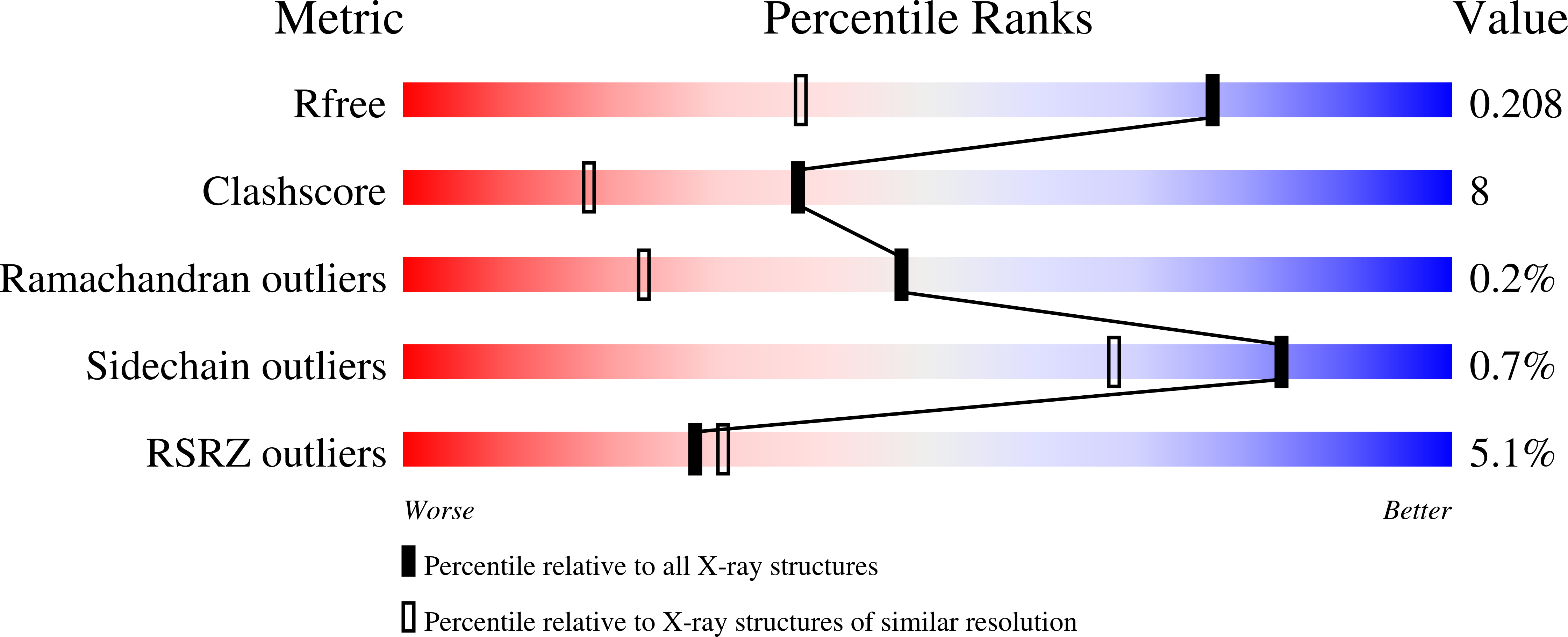
  • R-Value Free: 0.208 
  • R-Value Work: 0.179 

Starting Model: experimental
View more details

wwPDB Validation   3D Report Full Report


Ligand Structure Quality Assessment 

Created with Raphaël 2.3.0Worse 01 BetterLigand structure goodness of fit to experimental dataBest fitted 9JTClick on this verticalbar to view details

This is version 1.4 of the entry. See complete history


Literature

Crystallization and structure of ebselen bound to Cys141 of human inositol monophosphatase.

Fenn, G.D.Waller-Evans, H.Atack, J.R.Bax, B.D.

(2020) Acta Crystallogr F Struct Biol Commun 76: 469-476

  • DOI: https://doi.org/10.1107/S2053230X20011310
  • Primary Citation of Related Structures:  
    6ZK0

  • PubMed Abstract: 

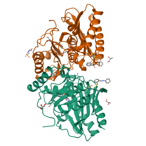
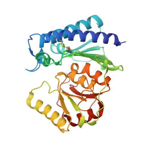
    Inositol monophosphatase (IMPase) is inhibited by lithium, which is the most efficacious treatment for bipolar disorder. Several therapies have been approved, or are going through clinical trials, aimed at the replacement of lithium in the treatment of bipolar disorder. One candidate small molecule is ebselen, a selenium-containing antioxidant, which has been demonstrated to produce lithium-like effects both in a murine model and in clinical trials. Here, the crystallization and the first structure of human IMPase covalently complexed with ebselen, a 1.47 Å resolution crystal structure (PDB entry 6zk0), are presented. In the complex with human IMPase, ebselen in a ring-opened conformation is covalently attached to Cys141, a residue located away from the active site. IMPase is a dimeric enzyme and in the crystal structure two adjacent dimers share four ebselen molecules, creating a tetramer with approximate 222 symmetry. In the crystal structure presented in this publication, the active site in the tetramer is still accessible, suggesting that ebselen may function as an allosteric inhibitor or may block the binding of partner proteins.


  • Organizational Affiliation

    Medicines Discovery Institute, School of Biosciences, Cardiff University, Cardiff CF10 3AT, United Kingdom.


Macromolecules
Find similar proteins by:  (by identity cutoff)  |  3D Structure
Entity ID: 1
MoleculeChains Sequence LengthOrganismDetailsImage
Inositol monophosphatase 1A [auth AAA],
B [auth BBB]
279Homo sapiensMutation(s): 0 
Gene Names: IMPA1IMPA
EC: 3.1.3.25 (PDB Primary Data), 3.1.3.94 (PDB Primary Data)
UniProt & NIH Common Fund Data Resources
Find proteins for P29218 (Homo sapiens)
Explore P29218 
Go to UniProtKB:  P29218
PHAROS:  P29218
GTEx:  ENSG00000133731 
Entity Groups  
Sequence Clusters30% Identity50% Identity70% Identity90% Identity95% Identity100% Identity
UniProt GroupP29218
Sequence Annotations
Expand
  • Reference Sequence
Small Molecules
Ligands 6 Unique
IDChains Name / Formula / InChI Key2D Diagram3D Interactions
9JT (Subject of Investigation/LOI)
Query on 9JT

Download Ideal Coordinates CCD File 
CA [auth BBB],
N [auth AAA]
N-phenyl-2-selanylbenzamide
C13 H11 N O Se
PVPUYGNPKBMXGO-UHFFFAOYSA-N
MES
Query on MES

Download Ideal Coordinates CCD File 
AA [auth BBB]2-(N-MORPHOLINO)-ETHANESULFONIC ACID
C6 H13 N O4 S
SXGZJKUKBWWHRA-UHFFFAOYSA-N
SO4
Query on SO4

Download Ideal Coordinates CCD File 
G [auth AAA],
Y [auth BBB]
SULFATE ION
O4 S
QAOWNCQODCNURD-UHFFFAOYSA-L
GOL
Query on GOL

Download Ideal Coordinates CCD File 
BA [auth BBB]
H [auth AAA]
I [auth AAA]
J [auth AAA]
K [auth AAA]
GLYCEROL
C3 H8 O3
PEDCQBHIVMGVHV-UHFFFAOYSA-N
MN
Query on MN

Download Ideal Coordinates CCD File 
D [auth AAA]
E [auth AAA]
T [auth BBB]
U [auth BBB]
V [auth BBB]
MANGANESE (II) ION
Mn
WAEMQWOKJMHJLA-UHFFFAOYSA-N
NA
Query on NA

Download Ideal Coordinates CCD File 
C [auth AAA]
DA [auth BBB]
F [auth AAA]
O [auth AAA]
P [auth AAA]
SODIUM ION
Na
FKNQFGJONOIPTF-UHFFFAOYSA-N
Experimental Data & Validation

Experimental Data

  • Method: X-RAY DIFFRACTION
  • Resolution: 1.47 Å
  • R-Value Free: 0.208 
  • R-Value Work: 0.179 
  • Space Group: P 32 2 1
Unit Cell:
Length ( Å )Angle ( ˚ )
a = 84.02α = 90
b = 84.02β = 90
c = 150.22γ = 120
Software Package:
Software NamePurpose
REFMACrefinement
XDSdata reduction
Aimlessdata scaling
REFMACphasing

Structure Validation

View Full Validation Report


Ligand Structure Quality Assessment 

Created with Raphaël 2.3.0Worse 01 BetterLigand structure goodness of fit to experimental dataBest fitted 9JTClick on this verticalbar to view details

Entry History 

Deposition Data

Revision History  (Full details and data files)

  • Version 1.0: 2020-09-23
    Type: Initial release
  • Version 1.1: 2020-10-14
    Changes: Database references
  • Version 1.2: 2023-04-12
    Changes: Database references, Derived calculations, Structure summary
  • Version 1.3: 2024-02-07
    Changes: Data collection, Refinement description
  • Version 1.4: 2024-10-16
    Changes: Structure summary