7JWV

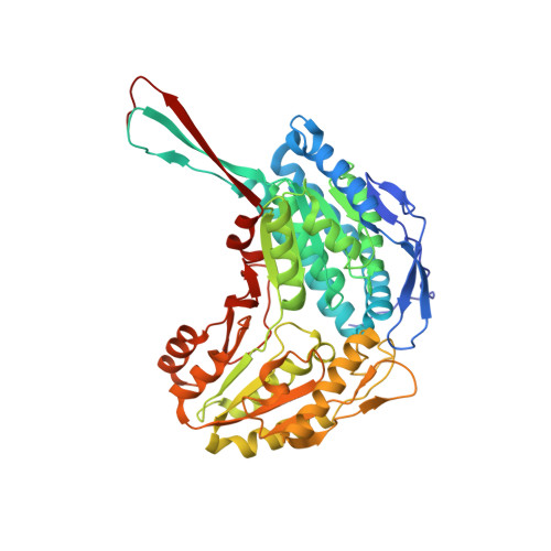
Crystal structure of human ALDH1A1 bound to compound (R)-28


Experimental Data Snapshot

  • Method: X-RAY DIFFRACTION
  • Resolution: 1.60 Å
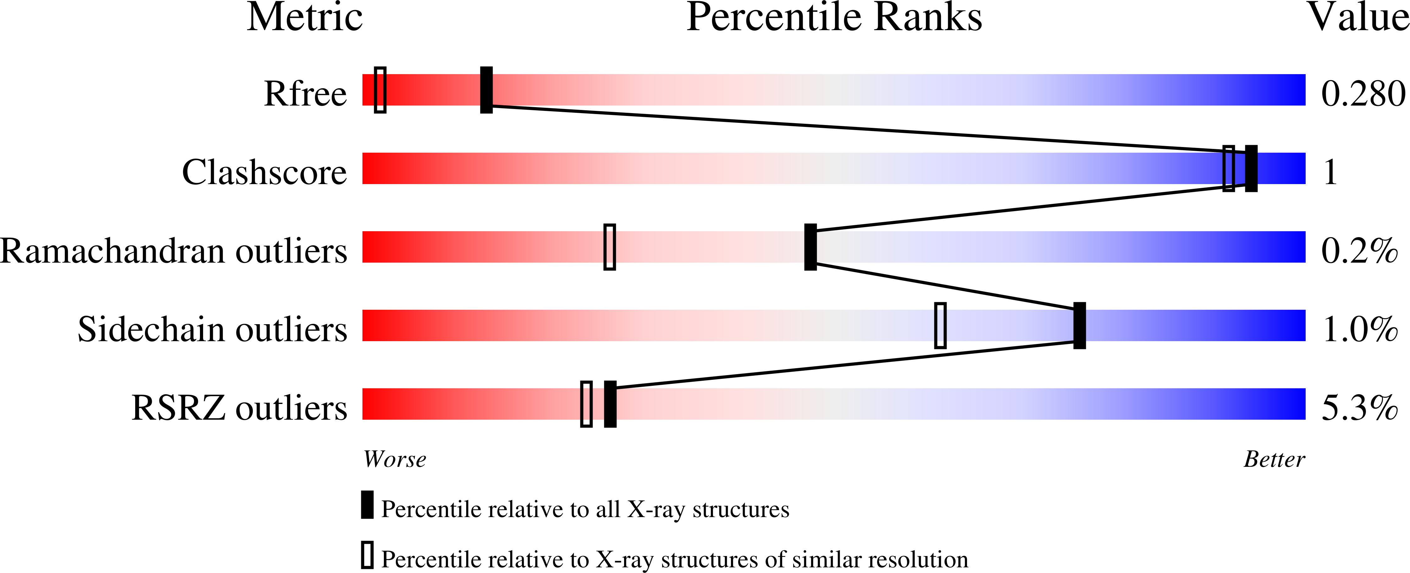
  • R-Value Free: 0.271 
  • R-Value Work: 0.235 
  • R-Value Observed: 0.236 

Starting Model: experimental
View more details

wwPDB Validation   3D Report Full Report


Ligand Structure Quality Assessment 


This is version 1.1 of the entry. See complete history


Literature

Development of 2,5-dihydro-4H-pyrazolo[3,4-d]pyrimidin-4-one inhibitors of aldehyde dehydrogenase 1A (ALDH1A) as potential adjuncts to ovarian cancer chemotherapy.

Huddle, B.C.Grimley, E.Chtcherbinine, M.Buchman, C.D.Takahashi, C.Debnath, B.McGonigal, S.C.Mao, S.Li, S.Felton, J.Pan, S.Wen, B.Sun, D.Neamati, N.Buckanovich, R.J.Hurley, T.D.Larsen, S.D.

(2020) Eur J Med Chem 211: 113060-113060

  • DOI: https://doi.org/10.1016/j.ejmech.2020.113060
  • Primary Citation of Related Structures:  
    7JWS, 7JWT, 7JWU, 7JWV, 7JWW

  • PubMed Abstract: 

    There is strong evidence that inhibition of one or more Aldehyde Dehydrogenase 1A (ALDH1A) isoforms may be beneficial in chemotherapy-resistant ovarian cancer and other tumor types. While many previous efforts have focused on development of ALDH1A1 selective inhibitors, the most deadly ovarian cancer subtype, high-grade serous (HGSOC), exhibits elevated expression of ALDH1A3. Herein, we report continued development of pan-ALDH1A inhibitors to assess whether broad spectrum ALDH1A inhibition is an effective adjunct to chemotherapy in this critical tumor subtype. Optimization of the CM39 scaffold, aided by metabolite ID and several new ALDH1A1 crystal structures, led to improved biochemical potencies, improved cellular ALDH inhibition in HGSOC cell lines, and substantial improvements in microsomal stability culminating in orally bioavailable compounds. We demonstrate that two compounds 68 and 69 are able to synergize with chemotherapy in a resistant cell line and patient-derived HGSOC tumor spheroids, indicating their suitability for future in vivo proof of concept experiments.


  • Organizational Affiliation

    Department of Medicinal Chemistry, College of Pharmacy, University of Michigan, Ann Arbor, MI, 48109, USA.


Macromolecules
Find similar proteins by:  (by identity cutoff)  |  3D Structure
Entity ID: 1
MoleculeChains Sequence LengthOrganismDetailsImage
Retinal dehydrogenase 1501Homo sapiensMutation(s): 1 
Gene Names: ALDH1A1ALDCALDH1PUMB1
EC: 1.2.1.19 (UniProt), 1.2.1.28 (UniProt), 1.2.1.36 (UniProt), 1.2.1.3 (UniProt)
UniProt & NIH Common Fund Data Resources
Find proteins for P00352 (Homo sapiens)
Explore P00352 
Go to UniProtKB:  P00352
PHAROS:  P00352
GTEx:  ENSG00000165092 
Entity Groups  
Sequence Clusters30% Identity50% Identity70% Identity90% Identity95% Identity100% Identity
UniProt GroupP00352
Sequence Annotations
Expand
  • Reference Sequence
Small Molecules
Binding Affinity Annotations 
IDSourceBinding Affinity
VMJ BindingDB:  7JWV IC50: 600 (nM) from 1 assay(s)
Experimental Data & Validation

Experimental Data

  • Method: X-RAY DIFFRACTION
  • Resolution: 1.60 Å
  • R-Value Free: 0.271 
  • R-Value Work: 0.235 
  • R-Value Observed: 0.236 
  • Space Group: P 4 2 2
Unit Cell:
Length ( Å )Angle ( ˚ )
a = 109.194α = 90
b = 109.194β = 90
c = 83.174γ = 90
Software Package:
Software NamePurpose
HKL-2000data reduction
HKL-2000data scaling
PHASERphasing
REFMACrefinement
PDB_EXTRACTdata extraction

Structure Validation

View Full Validation Report


Ligand Structure Quality Assessment 


Entry History & Funding Information

Deposition Data


Funding OrganizationLocationGrant Number
National Institutes of Health/National Cancer Institute (NIH/NCI)United StatesR01CA214567

Revision History  (Full details and data files)

  • Version 1.0: 2020-12-30
    Type: Initial release
  • Version 1.1: 2023-10-18
    Changes: Advisory, Data collection, Database references, Refinement description