8A1H | pdb_00008a1h

Bacterial 6-4 photolyase from Vibrio cholerase


Experimental Data Snapshot

  • Method: X-RAY DIFFRACTION
  • Resolution: 1.65 Å
  • R-Value Free: 
    0.136 (Depositor), 0.140 (DCC) 
  • R-Value Work: 
    0.119 (Depositor), 0.120 (DCC) 
  • R-Value Observed: 
    0.119 (Depositor) 

Starting Model: experimental
View more details

wwPDB Validation   3D Report Full Report


Ligand Structure Quality Assessment 

Created with Raphaël 2.3.0Worse 01 BetterLigand structure goodness of fit to experimental dataBest fitted FADClick on this verticalbar to view details

This is version 1.3 of the entry. See complete history


Literature

Structural and Functional Analysis of a Prokaryotic (6-4) Photolyase from the Aquatic Pathogen Vibrio Cholerae † .

Emmerich, H.J.Schneider, L.Essen, L.O.

(2023) Photochem Photobiol 99: 1248-1257

  • DOI: https://doi.org/10.1111/php.13783
  • Primary Citation of Related Structures:  
    8A1H

  • PubMed Abstract: 

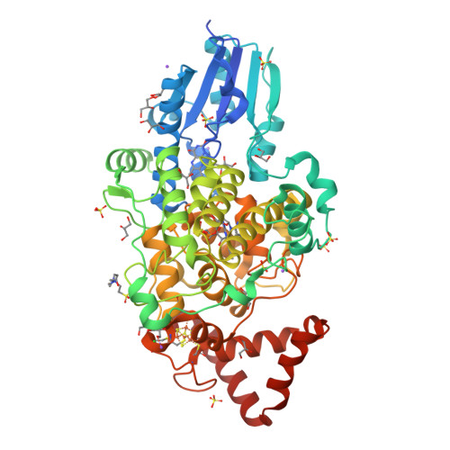
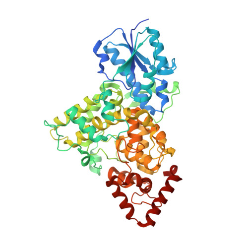
    Photolyases are flavoproteins, which are able to repair UV-induced DNA lesions in a light-dependent manner. According to their substrate, they can be distinguished as CPD- and (6-4) photolyases. While CPD-photolyases repair the predominantly occurring cyclobutane pyrimidine dimer lesion, (6-4) photolyases catalyze the repair of the less prominent (6-4) photoproduct. The subgroup of prokaryotic (6-4) photolyases/FeS-BCP is one of the most ancient types of flavoproteins in the ubiquitously occurring photolyase & cryptochrome superfamily (PCSf). In contrast to canonical photolyases, prokaryotic (6-4) photolyases possess a few particular characteristics, including a lumazine derivative as antenna chromophore besides the catalytically essential flavin adenine dinucleotide as well as an elongated linker region between the N-terminal α/β-domain and the C-terminal all-α-helical domain. Furthermore, they can harbor an additional short subdomain, located at the C-terminus, with a binding site for a [4Fe-4S] cluster. So far, two crystal structures of prokaryotic (6-4) photolyases have been reported. Within this study, we present the high-resolution structure of the prokaryotic (6-4) photolyase from Vibrio cholerae and its spectroscopic characterization in terms of in vitro photoreduction and DNA-repair activity.


  • Organizational Affiliation

    Unit for Structural Biochemistry, Department of Chemistry, Philipps University Marburg, Marburg, Germany.


Macromolecules
Find similar proteins by:  (by identity cutoff)  |  3D Structure
Entity ID: 1
MoleculeChains Sequence LengthOrganismDetailsImage
6-4 photolyase (FeS-BCP, CryPro)536Vibrio choleraeMutation(s): 0 
Gene Names: VC_A0809
UniProt
Find proteins for Q9KLD7 (Vibrio cholerae serotype O1 (strain ATCC 39315 / El Tor Inaba N16961))
Explore Q9KLD7 
Go to UniProtKB:  Q9KLD7
Entity Groups  
Sequence Clusters30% Identity50% Identity70% Identity90% Identity95% Identity100% Identity
UniProt GroupQ9KLD7
Sequence Annotations
Expand
  • Reference Sequence
Small Molecules
Ligands 8 Unique
IDChains Name / Formula / InChI Key2D Diagram3D Interactions
FAD (Subject of Investigation/LOI)
Query on FAD

Download Ideal Coordinates CCD File 
B [auth A]FLAVIN-ADENINE DINUCLEOTIDE
C27 H33 N9 O15 P2
VWWQXMAJTJZDQX-UYBVJOGSSA-N
SF4
Query on SF4

Download Ideal Coordinates CCD File 
D [auth A]IRON/SULFUR CLUSTER
Fe4 S4
LJBDFODJNLIPKO-UHFFFAOYSA-N
DLZ
Query on DLZ

Download Ideal Coordinates CCD File 
C [auth A]1-deoxy-1-(6,7-dimethyl-2,4-dioxo-3,4-dihydropteridin-8(2H)-yl)-D-ribitol
C13 H18 N4 O6
SXDXRJZUAJBNFL-XKSSXDPKSA-N
EPE
Query on EPE

Download Ideal Coordinates CCD File 
E [auth A]4-(2-HYDROXYETHYL)-1-PIPERAZINE ETHANESULFONIC ACID
C8 H18 N2 O4 S
JKMHFZQWWAIEOD-UHFFFAOYSA-N
SO4
Query on SO4

Download Ideal Coordinates CCD File 
Q [auth A]
R [auth A]
S [auth A]
T [auth A]
U [auth A]
SULFATE ION
O4 S
QAOWNCQODCNURD-UHFFFAOYSA-L
GOL
Query on GOL

Download Ideal Coordinates CCD File 
F [auth A]
G [auth A]
H [auth A]
I [auth A]
J [auth A]
GLYCEROL
C3 H8 O3
PEDCQBHIVMGVHV-UHFFFAOYSA-N
CL
Query on CL

Download Ideal Coordinates CCD File 
DA [auth A]CHLORIDE ION
Cl
VEXZGXHMUGYJMC-UHFFFAOYSA-M
NA
Query on NA

Download Ideal Coordinates CCD File 
AA [auth A]
BA [auth A]
CA [auth A]
X [auth A]
Y [auth A]
SODIUM ION
Na
FKNQFGJONOIPTF-UHFFFAOYSA-N
Experimental Data & Validation

Experimental Data

  • Method: X-RAY DIFFRACTION
  • Resolution: 1.65 Å
  • R-Value Free:  0.136 (Depositor), 0.140 (DCC) 
  • R-Value Work:  0.119 (Depositor), 0.120 (DCC) 
  • R-Value Observed: 0.119 (Depositor) 
Space Group: P 64 2 2
Unit Cell:
Length ( Å )Angle ( ˚ )
a = 199.687α = 90
b = 199.687β = 90
c = 76.996γ = 120
Software Package:
Software NamePurpose
PHENIXphasing
PHENIXrefinement
XDSdata reduction
Aimlessdata scaling

Structure Validation

View Full Validation Report


Ligand Structure Quality Assessment 

Created with Raphaël 2.3.0Worse 01 BetterLigand structure goodness of fit to experimental dataBest fitted FADClick on this verticalbar to view details

Entry History & Funding Information

Deposition Data


Funding OrganizationLocationGrant Number
German Research Foundation (DFG)GermanyES152/18

Revision History  (Full details and data files)

  • Version 1.0: 2023-02-08
    Type: Initial release
  • Version 1.1: 2023-02-22
    Changes: Database references
  • Version 1.2: 2023-10-04
    Changes: Data collection, Database references
  • Version 1.3: 2024-02-07
    Changes: Refinement description