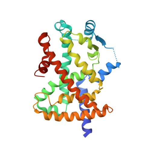
8HHP

Crystal structure of PPARg-LBD complexed with three partial agonists, one nTZDpa and two LT175


Experimental Data Snapshot

  • Method: X-RAY DIFFRACTION
  • Resolution: 2.45 Å
  • R-Value Free: 0.286 
  • R-Value Work: 0.214 
  • R-Value Observed: 0.221 

wwPDB Validation   3D Report Full Report


Ligand Structure Quality Assessment 


This is version 1.1 of the entry. See complete history


Literature

Covalent Modifier Discovery Using Hydrogen/Deuterium Exchange-Mass Spectrometry.

Kojima, H.Yanagi, R.Higuchi, E.Yoshizawa, M.Shimodaira, T.Kumagai, M.Kyoya, T.Sekine, M.Egawa, D.Ohashi, N.Ishida, H.Yamamoto, K.Itoh, T.

(2023) J Med Chem 66: 4827-4839

  • DOI: https://doi.org/10.1021/acs.jmedchem.2c01986
  • Primary Citation of Related Structures:  
    8HHP, 8HHQ

  • PubMed Abstract: 

    Covalent ligands are generally filtered out of chemical libraries used for high-throughput screening, because electrophilic functional groups are considered to be pan-assay interference compounds (PAINS). Therefore, screening strategies that can distinguish true covalent ligands from PAINS are required. Hydrogen/deuterium-exchange mass spectrometry (HDX-MS) is a powerful tool for evaluating protein stability. Here, we report a covalent modifier screening approach using HDX-MS. In this study, HDX-MS was used to classify peroxisome proliferator-activated receptor γ (PPARγ) and vitamin D receptor ligands. HDX-MS could discriminate the strength of ligand-protein interactions. Our HDX-MS screening method identified LT175 and nTZDpa, which can bind concurrently to the PPARγ ligand-binding domain (PPARγ-LBD) with synergistic activation. Furthermore, iodoacetic acid was identified as a novel covalent modifier that stabilizes the PPARγ-LBD.


  • Organizational Affiliation

    Laboratory of Drug Design and Medicinal Chemistry, Showa Pharmaceutical University, 3-3165 Higashi-Tamagawagakuen, Machida, Tokyo 194-8543, Japan.


Macromolecules
Find similar proteins by:  (by identity cutoff)  |  3D Structure
Entity ID: 1
MoleculeChains Sequence LengthOrganismDetailsImage
Peroxisome proliferator-activated receptor gamma276Homo sapiensMutation(s): 0 
Gene Names: PPARG
UniProt & NIH Common Fund Data Resources
Find proteins for P37231 (Homo sapiens)
Explore P37231 
Go to UniProtKB:  P37231
PHAROS:  P37231
GTEx:  ENSG00000132170 
Entity Groups  
Sequence Clusters30% Identity50% Identity70% Identity90% Identity95% Identity100% Identity
UniProt GroupP37231
Sequence Annotations
Expand
  • Reference Sequence
Small Molecules
Binding Affinity Annotations 
IDSourceBinding Affinity
NZA BindingDB:  8HHP Kd: 29 (nM) from 1 assay(s)
IC50: 26 (nM) from 1 assay(s)
EC50: min: 55, max: 57 (nM) from 2 assay(s)
LRG BindingDB:  8HHP EC50: min: 480, max: 562 (nM) from 5 assay(s)
Experimental Data & Validation

Experimental Data

  • Method: X-RAY DIFFRACTION
  • Resolution: 2.45 Å
  • R-Value Free: 0.286 
  • R-Value Work: 0.214 
  • R-Value Observed: 0.221 
  • Space Group: P 43 21 2
Unit Cell:
Length ( Å )Angle ( ˚ )
a = 60.5α = 90
b = 60.5β = 90
c = 162.26γ = 90
Software Package:
Software NamePurpose
PHENIXrefinement
iMOSFLMdata reduction
Aimlessdata scaling
PHASERphasing

Structure Validation

View Full Validation Report


Ligand Structure Quality Assessment 


Entry History & Funding Information

Deposition Data


Funding OrganizationLocationGrant Number
Japan Agency for Medical Research and Development (AMED)Japan--

Revision History  (Full details and data files)

  • Version 1.0: 2023-11-22
    Type: Initial release
  • Version 1.1: 2024-05-22
    Changes: Database references