8OWZ

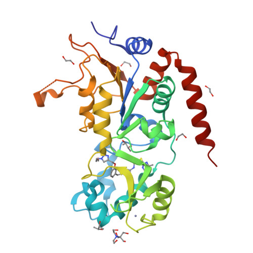
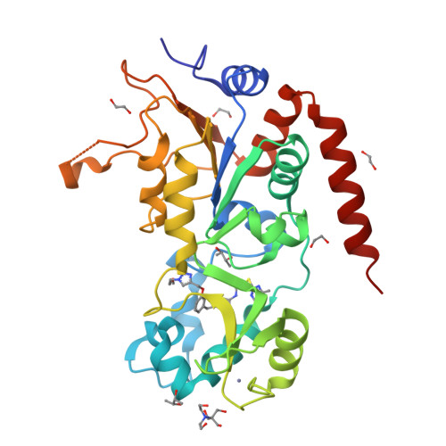
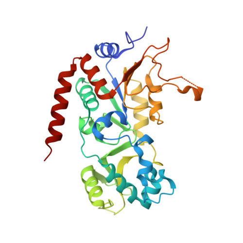
Crystal structure of human Sirt2 in complex with a triazole-based SirReal


Experimental Data Snapshot

  • Method: X-RAY DIFFRACTION
  • Resolution: 1.65 Å
  • R-Value Free: 0.198 
  • R-Value Work: 0.168 
  • R-Value Observed: 0.170 

Starting Model: experimental
View more details

wwPDB Validation   3D Report Full Report


Ligand Structure Quality Assessment 


This is version 1.1 of the entry. See complete history


Literature

Development of First-in-Class Dual Sirt2/HDAC6 Inhibitors as Molecular Tools for Dual Inhibition of Tubulin Deacetylation.

Sinatra, L.Vogelmann, A.Friedrich, F.Tararina, M.A.Neuwirt, E.Colcerasa, A.Konig, P.Toy, L.Yesiloglu, T.Z.Hilscher, S.Gaitzsch, L.Papenkordt, N.Zhai, S.Zhang, L.Romier, C.Einsle, O.Sippl, W.Schutkowski, M.Gross, O.Bendas, G.Christianson, D.W.Hansen, F.K.Jung, M.Schiedel, M.

(2023) J Med Chem 66: 14787-14814

  • DOI: https://doi.org/10.1021/acs.jmedchem.3c01385
  • Primary Citation of Related Structures:  
    8G1Z, 8G20, 8OWZ

  • PubMed Abstract: 

    Dysregulation of both tubulin deacetylases sirtuin 2 (Sirt2) and the histone deacetylase 6 (HDAC6) has been associated with the pathogenesis of cancer and neurodegeneration, thus making these two enzymes promising targets for pharmaceutical intervention. Herein, we report the design, synthesis, and biological characterization of the first-in-class dual Sirt2/HDAC6 inhibitors as molecular tools for dual inhibition of tubulin deacetylation. Using biochemical in vitro assays and cell-based methods for target engagement, we identified Mz325 ( 33 ) as a potent and selective inhibitor of both target enzymes. Inhibition of both targets was further confirmed by X-ray crystal structures of Sirt2 and HDAC6 in complex with building blocks of 33 . In ovarian cancer cells, 33 evoked enhanced effects on cell viability compared to single or combination treatment with the unconjugated Sirt2 and HDAC6 inhibitors. Thus, our dual Sirt2/HDAC6 inhibitors are important new tools to study the consequences and the therapeutic potential of dual inhibition of tubulin deacetylation.


  • Organizational Affiliation

    Institute for Drug Discovery, Medical Faculty, Leipzig University, Brüderstraße 34, 04103 Leipzig, Germany.


Macromolecules
Find similar proteins by:  (by identity cutoff)  |  3D Structure
Entity ID: 1
MoleculeChains Sequence LengthOrganismDetailsImage
NAD-dependent protein deacetylase sirtuin-2304Homo sapiensMutation(s): 0 
Gene Names: SIRT2SIR2LSIR2L2
EC: 3.5.1 (PDB Primary Data), 2.3.1 (UniProt), 2.3.1.286 (UniProt)
UniProt & NIH Common Fund Data Resources
Find proteins for Q8IXJ6 (Homo sapiens)
Explore Q8IXJ6 
Go to UniProtKB:  Q8IXJ6
PHAROS:  Q8IXJ6
GTEx:  ENSG00000068903 
Entity Groups  
Sequence Clusters30% Identity50% Identity70% Identity90% Identity95% Identity100% Identity
UniProt GroupQ8IXJ6
Sequence Annotations
Expand
  • Reference Sequence
Small Molecules
Ligands 6 Unique
IDChains Name / Formula / InChI Key2D Diagram3D Interactions
KZU (Subject of Investigation/LOI)
Query on KZU

Download Ideal Coordinates CCD File 
B [auth A]2-(4,6-dimethylpyrimidin-2-yl)sulfanyl-N-[5-[[3-[[1-(2-methoxyethyl)-1,2,3-triazol-4-yl]methoxy]phenyl]methyl]-1,3-thiazol-2-yl]ethanamide
C24 H27 N7 O3 S2
MAUGCLAAQLDMTD-UHFFFAOYSA-N
BTB
Query on BTB

Download Ideal Coordinates CCD File 
D [auth A]2-[BIS-(2-HYDROXY-ETHYL)-AMINO]-2-HYDROXYMETHYL-PROPANE-1,3-DIOL
C8 H19 N O5
OWMVSZAMULFTJU-UHFFFAOYSA-N
BU3
Query on BU3

Download Ideal Coordinates CCD File 
E [auth A],
F [auth A]
(R,R)-2,3-BUTANEDIOL
C4 H10 O2
OWBTYPJTUOEWEK-QWWZWVQMSA-N
ZN
Query on ZN

Download Ideal Coordinates CCD File 
J [auth A]ZINC ION
Zn
PTFCDOFLOPIGGS-UHFFFAOYSA-N
EDO
Query on EDO

Download Ideal Coordinates CCD File 
C [auth A],
G [auth A],
H [auth A],
I [auth A]
1,2-ETHANEDIOL
C2 H6 O2
LYCAIKOWRPUZTN-UHFFFAOYSA-N
CL
Query on CL

Download Ideal Coordinates CCD File 
K [auth A]CHLORIDE ION
Cl
VEXZGXHMUGYJMC-UHFFFAOYSA-M
Binding Affinity Annotations 
IDSourceBinding Affinity
KZU BindingDB:  8OWZ IC50: min: 118, max: 2500 (nM) from 3 assay(s)
Experimental Data & Validation

Experimental Data

  • Method: X-RAY DIFFRACTION
  • Resolution: 1.65 Å
  • R-Value Free: 0.198 
  • R-Value Work: 0.168 
  • R-Value Observed: 0.170 
  • Space Group: P 1 21 1
Unit Cell:
Length ( Å )Angle ( ˚ )
a = 35.81α = 90
b = 73.463β = 95.34
c = 55.333γ = 90
Software Package:
Software NamePurpose
XDSdata reduction
Aimlessdata scaling
PHASERphasing
PHENIXrefinement

Structure Validation

View Full Validation Report


Ligand Structure Quality Assessment 


Entry History & Funding Information

Deposition Data


Funding OrganizationLocationGrant Number
German Research Foundation (DFG)GermanySFB 992
German Research Foundation (DFG)Germany295/18-1

Revision History  (Full details and data files)

  • Version 1.0: 2023-11-15
    Type: Initial release
  • Version 1.1: 2023-11-22
    Changes: Database references