Crystal structure of alcohol dehydrogenase/ketoreductase variant from Thermus thermophilus apo form
Delgado-Arciniega, E., Bodescu, V., Rozeboom, H.J., Janssen, D.B., Wijma, H.J.To be published.
Experimental Data Snapshot
Starting Model: experimental
View more details
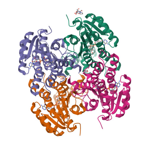
Entity ID: 1 | |||||
|---|---|---|---|---|---|
| Molecule | Chains | Sequence Length | Organism | Details | Image |
| Oxidoreductase, short-chain dehydrogenase/reductase family | 264 | Thermus thermophilus HB8 | Mutation(s): 9 Gene Names: TTHA0369 | 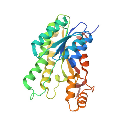 | |
UniProt | |||||
Find proteins for Q5SLC4 (Thermus thermophilus (strain ATCC 27634 / DSM 579 / HB8)) Explore Q5SLC4 Go to UniProtKB: Q5SLC4 | |||||
Entity Groups | |||||
| Sequence Clusters | 30% Identity50% Identity70% Identity90% Identity95% Identity100% Identity | ||||
| UniProt Group | Q5SLC4 | ||||
Sequence AnnotationsExpand | |||||
| |||||
| Ligands 3 Unique | |||||
|---|---|---|---|---|---|
| ID | Chains | Name / Formula / InChI Key | 2D Diagram | 3D Interactions | |
| NAD (Subject of Investigation/LOI) Query on NAD | E [auth A], G [auth B], J [auth C], L [auth D] | NICOTINAMIDE-ADENINE-DINUCLEOTIDE C21 H27 N7 O14 P2 BAWFJGJZGIEFAR-NNYOXOHSSA-N |  | ||
| B3P Query on B3P | F [auth A] | 2-[3-(2-HYDROXY-1,1-DIHYDROXYMETHYL-ETHYLAMINO)-PROPYLAMINO]-2-HYDROXYMETHYL-PROPANE-1,3-DIOL C11 H26 N2 O6 HHKZCCWKTZRCCL-UHFFFAOYSA-N |  | ||
| GOL Query on GOL | H [auth B], I [auth C], K [auth D] | GLYCEROL C3 H8 O3 PEDCQBHIVMGVHV-UHFFFAOYSA-N |  | ||
| Length ( Å ) | Angle ( ˚ ) |
|---|---|
| a = 70.981 | α = 90 |
| b = 92.937 | β = 107.87 |
| c = 79.601 | γ = 90 |
| Software Name | Purpose |
|---|---|
| REFMAC | refinement |
| Aimless | data scaling |
| XDS | data reduction |
| PHASER | phasing |
| Funding Organization | Location | Grant Number |
|---|---|---|
| Not funded | -- |
RCSB PDB is hosted by

RCSB PDB is a member of the