1AYL | pdb_00001ayl

PHOSPHOENOLPYRUVATE CARBOXYKINASE


Experimental Data Snapshot

  • Method: X-RAY DIFFRACTION
  • Resolution: 1.80 Å
  • R-Value Free: 
    0.234 (Depositor) 
  • R-Value Work: 
    0.195 (Depositor) 
  • R-Value Observed: 
    0.195 (Depositor) 

wwPDB Validation   3D Report Full Report


This is version 1.5 of the entry. See complete history


Literature

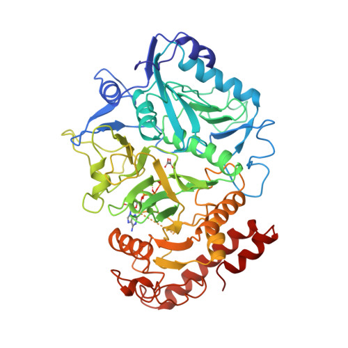
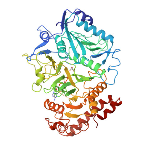
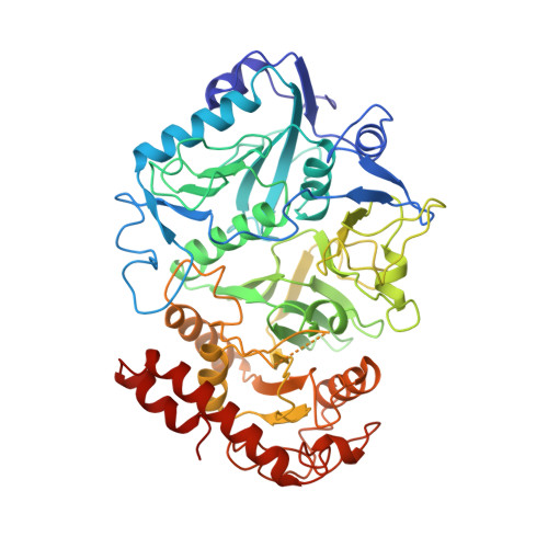
Snapshot of an enzyme reaction intermediate in the structure of the ATP-Mg2+-oxalate ternary complex of Escherichia coli PEP carboxykinase.

Tari, L.W.Matte, A.Pugazhenthi, U.Goldie, H.Delbaere, L.T.

(1996) Nat Struct Biol 3: 355-363

  • DOI: https://doi.org/10.1038/nsb0496-355
  • Primary Citation of Related Structures:  
    1AYL

  • PubMed Abstract: 

    We report the 1.8 A crystal structure of adenosine triphosphate (ATP)-magnesium-oxalate bound phosphoenolpyruvate carboxykinase (PCK) from Escherichia coli. ATP binding induces a 20 degree hinge-like rotation of the N- and C-terminal domains which closes the active-site cleft. PCK possesses a novel nucleotide-binding fold, particularly in the adenine-binding region, where the formation of a cis backbone torsion angle in a loop glycine residue promotes intimate contacts between the adenine-binding loop and adenine, while stabilizing a syn conformation of the base. This complex represents a reaction intermediate analogue along the pathway of the conversion of oxaloacetate to phosphoenolpyruvate, and provides insight into the mechanistic details of the chemical reaction catalysed by this enzyme.


  • Organizational Affiliation

    Department of Biochemistry, University of Saskatchewan, Saskatoon, Saskatchewan, Canada.


Macromolecules
Find similar proteins by:  (by identity cutoff)  |  3D Structure
Entity ID: 1
MoleculeChains Sequence LengthOrganismDetailsImage
PHOSPHOENOLPYRUVATE CARBOXYKINASE541Escherichia coli K-12Mutation(s): 0 
EC: 4.1.1.49
UniProt
Find proteins for P22259 (Escherichia coli (strain K12))
Explore P22259 
Go to UniProtKB:  P22259
Entity Groups  
Sequence Clusters30% Identity50% Identity70% Identity90% Identity95% Identity100% Identity
UniProt GroupP22259
Sequence Annotations
Expand
  • Reference Sequence
Small Molecules
Ligands 3 Unique
IDChains Name / Formula / InChI Key2D Diagram3D Interactions
ATP
Query on ATP

Download Ideal Coordinates CCD File 
D [auth A]ADENOSINE-5'-TRIPHOSPHATE
C10 H16 N5 O13 P3
ZKHQWZAMYRWXGA-KQYNXXCUSA-N
OXL
Query on OXL

Download Ideal Coordinates CCD File 
B [auth A]OXALATE ION
C2 O4
MUBZPKHOEPUJKR-UHFFFAOYSA-L
MG
Query on MG

Download Ideal Coordinates CCD File 
C [auth A]MAGNESIUM ION
Mg
JLVVSXFLKOJNIY-UHFFFAOYSA-N
Experimental Data & Validation

Experimental Data

  • Method: X-RAY DIFFRACTION
  • Resolution: 1.80 Å
  • R-Value Free:  0.234 (Depositor) 
  • R-Value Work:  0.195 (Depositor) 
  • R-Value Observed: 0.195 (Depositor) 
Space Group: C 1 2 1
Unit Cell:
Length ( Å )Angle ( ˚ )
a = 127.51α = 90
b = 96.69β = 96.63
c = 46.96γ = 90
Software Package:
Software NamePurpose
X-PLORrefinement
WEISdata reduction

Structure Validation

View Full Validation Report


Entry History 

Deposition Data

Revision History  (Full details and data files)

  • Version 1.0: 1997-01-11
    Type: Initial release
  • Version 1.1: 2008-03-24
    Changes: Version format compliance
  • Version 1.2: 2011-07-13
    Changes: Source and taxonomy, Version format compliance
  • Version 1.3: 2017-11-29
    Changes: Derived calculations, Other
  • Version 1.4: 2018-03-21
    Changes: Data collection
  • Version 1.5: 2024-02-07
    Changes: Data collection, Database references, Derived calculations