1T7D | pdb_00001t7d

Crystal structure of Escherichia coli type I signal peptidase in complex with a lipopeptide inhibitor


Experimental Data Snapshot

  • Method: X-RAY DIFFRACTION
  • Resolution: 2.47 Å
  • R-Value Free: 
    0.283 (Depositor), 0.280 (DCC) 
  • R-Value Work: 
    0.230 (Depositor), 0.230 (DCC) 
  • R-Value Observed: 
    0.230 (Depositor) 

Starting Model: experimental
View more details

wwPDB Validation   3D Report Full Report


Ligand Structure Quality Assessment 

Created with Raphaël 2.3.0Worse 01 BetterLigand structure goodness of fit to experimental dataBest fitted M12Click on this verticalbar to view details

This is version 2.0 of the entry. See complete history


Literature

Crystallographic and Biophysical Analysis of a Bacterial Signal Peptidase in Complex with a Lipopeptide Based Inhibitor.

Paetzel, M.Goodall, J.J.Kania, M.Dalbey, R.E.Page, M.G.P.

(2004) J Biological Chem 279: 30781

  • DOI: https://doi.org/10.1074/jbc.M401686200
  • Primary Citation of Related Structures:  
    1T7D

  • PubMed Abstract: 

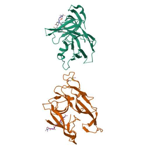
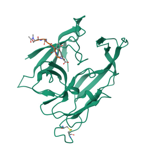
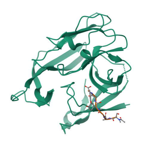
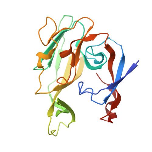
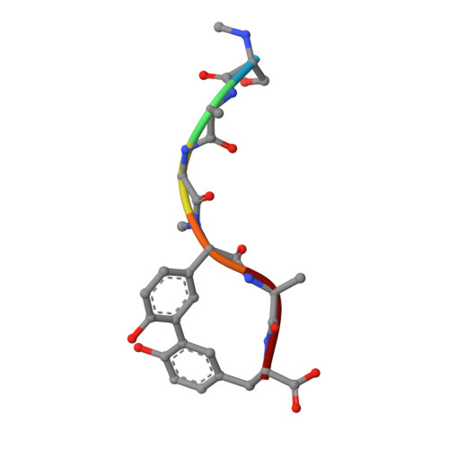
    We report here the crystallographic and biophysical analysis of a soluble, catalytically active fragment of the Escherichia coli type I signal peptidase (SPase Delta2-75) in complex with arylomycin A2. The 2.5-A resolution structure revealed that the inhibitor is positioned with its COOH-terminal carboxylate oxygen (O45) within hydrogen bonding distance of all the functional groups in the catalytic center of the enzyme (Ser90 O-gamma, Lys145 N-zeta, and Ser88 O-gamma) and that it makes beta-sheet type interactions with the beta-strands that line each side of the binding site. Ligand binding studies, calorimetry, fluorescence spectroscopy, and stopped-flow kinetics were also used to analyze the binding mode of this unique non-covalently bound inhibitor. The crystal structure was solved in the space group P4(3)2(1)2. A detailed comparison is made to the previously published acyl-enzyme inhibitor complex structure (space group: P2(1)2(1)2) and the apo-enzyme structure (space group: P4(1)2(1)2). Together this work provides insights into the binding of pre-protein substrates to signal peptidase and will prove helpful in the development of novel antibiotics.


  • Organizational Affiliation

    Department of Molecular Biology and Biochemistry, Simon Fraser University, Burnaby, British Columbia, V5A 1S6 Canada. mpaetze@sfu.ca


Macromolecules
Find similar proteins by:  (by identity cutoff)  |  3D Structure
Entity ID: 1
MoleculeChains Sequence LengthOrganismDetailsImage
SIGNAL PEPTIDASE I
A, B
250Escherichia coliMutation(s): 0 
Gene Names: LEPB
EC: 3.4.21.89
Membrane Entity: Yes 
UniProt
Find proteins for P00803 (Escherichia coli (strain K12))
Explore P00803 
Go to UniProtKB:  P00803
Entity Groups  
Sequence Clusters30% Identity50% Identity70% Identity90% Identity95% Identity100% Identity
UniProt GroupP00803
Sequence Annotations
Expand
  • Reference Sequence

Find similar proteins by:  Sequence   |   3D Structure  

Entity ID: 2
MoleculeChains Sequence LengthOrganismDetailsImage
ARYLOMYCIN A2
C, D
6Streptomyces sp.Mutation(s): 0 
Sequence Annotations
Expand
  • Reference Sequence
Small Molecules
Modified Residues  1 Unique
IDChains TypeFormula2D DiagramParent
5PG
Query on 5PG
C, D
L-PEPTIDE LINKINGC9 H11 N O3GLY
Biologically Interesting Molecules (External Reference) 1 Unique
Experimental Data & Validation

Experimental Data

  • Method: X-RAY DIFFRACTION
  • Resolution: 2.47 Å
  • R-Value Free:  0.283 (Depositor), 0.280 (DCC) 
  • R-Value Work:  0.230 (Depositor), 0.230 (DCC) 
  • R-Value Observed: 0.230 (Depositor) 
Space Group: P 43 21 2
Unit Cell:
Length ( Å )Angle ( ˚ )
a = 69.609α = 90
b = 69.609β = 90
c = 258.466γ = 90
Software Package:
Software NamePurpose
AMoREphasing
CNSrefinement
DENZOdata reduction
SCALEPACKdata scaling

Structure Validation

View Full Validation Report


Ligand Structure Quality Assessment 

Created with Raphaël 2.3.0Worse 01 BetterLigand structure goodness of fit to experimental dataBest fitted M12Click on this verticalbar to view details

Entry History 

Deposition Data

Revision History  (Full details and data files)

  • Version 1.0: 2004-07-13
    Type: Initial release
  • Version 1.1: 2011-06-14
    Changes: Version format compliance
  • Version 1.2: 2011-07-13
    Changes: Version format compliance
  • Version 1.3: 2011-07-27
    Changes: Atomic model, Database references, Derived calculations, Non-polymer description, Structure summary
  • Version 1.4: 2012-12-12
    Changes: Other
  • Version 2.0: 2023-08-23
    Changes: Data collection, Database references, Derived calculations, Polymer sequence, Refinement description