3FCO | pdb_00003fco

Crystal Structure of 11beta-Hydroxysteroid Dehydrogenase 1 (11b-HSD1) in Complex with Benzamide Inhibitor


Experimental Data Snapshot

  • Method: X-RAY DIFFRACTION
  • Resolution: 2.65 Å
  • R-Value Free: 
    0.254 (Depositor), 0.250 (DCC) 
  • R-Value Work: 
    0.222 (Depositor), 0.220 (DCC) 
  • R-Value Observed: 
    0.224 (Depositor) 

wwPDB Validation   3D Report Full Report


Ligand Structure Quality Assessment 

Created with Raphaël 2.3.0Worse 01 BetterLigand structure goodness of fit to experimental dataBest fitted NAPClick on this verticalbar to view detailsBest fitted IIGClick on this verticalbar to view details

This is version 1.3 of the entry. See complete history


Literature

Optimization of novel di-substituted cyclohexylbenzamide derivatives as potent 11 beta-HSD1 inhibitors.

McMinn, D.L.Rew, Y.Sudom, A.Caille, S.Degraffenreid, M.He, X.Hungate, R.Jiang, B.Jaen, J.Julian, L.D.Kaizerman, J.Novak, P.Sun, D.Tu, H.Ursu, S.Walker, N.P.Yan, X.Ye, Q.Wang, Z.Powers, J.P.

(2009) Bioorg Med Chem Lett 19: 1446-1450

  • DOI: https://doi.org/10.1016/j.bmcl.2009.01.026
  • Primary Citation of Related Structures:  
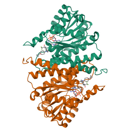
    3FCO

  • PubMed Abstract: 

    Novel 4,4-disubstituted cyclohexylbenzamide inhibitors of 11beta-HSD1 were optimized to account for liabilities relating to in vitro pharmacokinetics, cytotoxicity and protein-related shifts in potency. A representative compound showing favorable in vivo pharmacokinetics was found to be an efficacious inhibitor of 11beta-HSD1 in a rat pharmacodynamic model (ED(50)=10mg/kg).


  • Organizational Affiliation

    Amgen, Inc., 1120 Veterans Boulevard, South San Francisco, CA 94080, USA.


Macromolecules
Find similar proteins by:  (by identity cutoff)  |  3D Structure
Entity ID: 1
MoleculeChains Sequence LengthOrganismDetailsImage
Corticosteroid 11-beta-dehydrogenase isozyme 1
A, B
286Homo sapiensMutation(s): 1 
Gene Names: HSD11B1HSD11HSD11L
EC: 1.1.1.146 (PDB Primary Data), 1.1.1.201 (UniProt)
UniProt & NIH Common Fund Data Resources
Find proteins for P28845 (Homo sapiens)
Explore P28845 
Go to UniProtKB:  P28845
PHAROS:  P28845
GTEx:  ENSG00000117594 
Entity Groups  
Sequence Clusters30% Identity50% Identity70% Identity90% Identity95% Identity100% Identity
UniProt GroupP28845
Sequence Annotations
Expand
  • Reference Sequence
Small Molecules
Binding Affinity Annotations 
IDSourceBinding Affinity
IIG BindingDB:  3FCO IC50: min: 14, max: 121 (nM) from 4 assay(s)
Experimental Data & Validation

Experimental Data

  • Method: X-RAY DIFFRACTION
  • Resolution: 2.65 Å
  • R-Value Free:  0.254 (Depositor), 0.250 (DCC) 
  • R-Value Work:  0.222 (Depositor), 0.220 (DCC) 
  • R-Value Observed: 0.224 (Depositor) 
Space Group: P 31 2 1
Unit Cell:
Length ( Å )Angle ( ˚ )
a = 109.185α = 90
b = 109.185β = 90
c = 135.869γ = 120
Software Package:
Software NamePurpose
MOSFLMdata reduction
SCALAdata scaling
MOLREPphasing
REFMACrefinement
PDB_EXTRACTdata extraction
BOSdata collection

Structure Validation

View Full Validation Report


Ligand Structure Quality Assessment 

Created with Raphaël 2.3.0Worse 01 BetterLigand structure goodness of fit to experimental dataBest fitted NAPClick on this verticalbar to view detailsBest fitted IIGClick on this verticalbar to view details

Entry History 

Deposition Data

Revision History  (Full details and data files)

  • Version 1.0: 2009-12-01
    Type: Initial release
  • Version 1.1: 2011-07-13
    Changes: Version format compliance
  • Version 1.2: 2021-10-20
    Changes: Database references, Derived calculations
  • Version 1.3: 2023-12-27
    Changes: Data collection