3TPQ | pdb_00003tpq

Crystal structure of wild-type MAL RPEL domain in complex with five G-actins


Experimental Data Snapshot

  • Method: X-RAY DIFFRACTION
  • Resolution: 3.45 Å
  • R-Value Free: 
    0.273 (Depositor), 0.270 (DCC) 
  • R-Value Work: 
    0.224 (Depositor), 0.220 (DCC) 
  • R-Value Observed: 
    0.227 (Depositor) 

Starting Model: experimental
View more details

wwPDB Validation   3D Report Full Report


Ligand Structure Quality Assessment 

Created with Raphaël 2.3.0Worse 01 BetterLigand structure goodness of fit to experimental dataBest fitted ATPClick on this verticalbar to view details

This is version 1.2 of the entry. See complete history


Literature

Sensing actin dynamics: structural basis for G-actin-sensitive nuclear import of MAL

Hirano, H.Matsuura, Y.

(2011) Biochem Biophys Res Commun 414: 373-378

  • DOI: https://doi.org/10.1016/j.bbrc.2011.09.079
  • Primary Citation of Related Structures:  
    3TPM, 3TPO, 3TPQ

  • PubMed Abstract: 

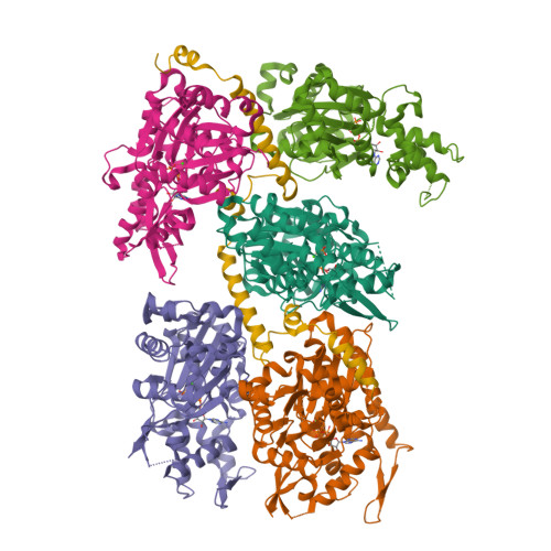
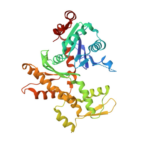
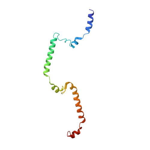
    The coordination of cytoskeletal actin dynamics with gene expression reprogramming is emerging as a crucial mechanism to control diverse cellular processes, including cell migration, differentiation and neuronal circuit assembly. The actin-binding transcriptional coactivator MAL (also known as MRTF-A/MKL1/BSAC) senses G-actin concentration and transduces Rho GTPase signals to serum response factor (SRF). MAL rapidly shuttles between the cytoplasm and the nucleus in unstimulated cells but Rho-induced depletion of G-actin leads to MAL nuclear accumulation and activation of transcription of SRF:MAL-target genes. Although the molecular and structural basis of actin-regulated nucleocytoplasmic shuttling of MAL is not understood fully, it is proposed that nuclear import of MAL is mediated by importin α/β heterodimer, and that G-actin competes with importin α/β for the binding to MAL. Here we present structural, biochemical and cell biological evidence that MAL has a classical bipartite nuclear localization signal (NLS) in the N-terminal 'RPEL' domain containing Arg-Pro-X-X-X-Glu-Leu (RPEL) motifs. The NLS residues of MAL adopt an extended conformation and bind along the surface groove of importin-α, interacting with the major- and minor-NLS binding sites. We also present a crystal structure of wild-type MAL RPEL domain in complex with five G-actins. Comparison of the importin-α- and actin-complexes revealed that the binding of G-actins to MAL is associated with folding of NLS residues into a helical conformation that is inappropriate for importin-α recognition.


  • Organizational Affiliation

    Structural Biology Research Center, Graduate School of Science, Nagoya University, Nagoya 464-8602, Japan.


Macromolecules
Find similar proteins by:  (by identity cutoff)  |  3D Structure
Entity ID: 1
MoleculeChains Sequence LengthOrganismDetailsImage
Actin, alpha skeletal muscle
A, B, C, D, E
375Oryctolagus cuniculusMutation(s): 0 
EC: 3.6.4
UniProt
Find proteins for P68135 (Oryctolagus cuniculus)
Explore P68135 
Go to UniProtKB:  P68135
Entity Groups  
Sequence Clusters30% Identity50% Identity70% Identity90% Identity95% Identity100% Identity
UniProt GroupP68135
Sequence Annotations
Expand
  • Reference Sequence
Find similar proteins by:  (by identity cutoff)  |  3D Structure
Entity ID: 2
MoleculeChains Sequence LengthOrganismDetailsImage
MALF [auth M]117Mus musculusMutation(s): 0 
Gene Names: Mkl1
Entity Groups  
Sequence Clusters30% Identity50% Identity70% Identity90% Identity95% Identity100% Identity
Sequence Annotations
Expand
  • Reference Sequence
Small Molecules
Ligands 2 Unique
IDChains Name / Formula / InChI Key2D Diagram3D Interactions
ATP
Query on ATP

Download Ideal Coordinates CCD File 
G [auth A],
I [auth B],
K [auth C],
M [auth D],
O [auth E]
ADENOSINE-5'-TRIPHOSPHATE
C10 H16 N5 O13 P3
ZKHQWZAMYRWXGA-KQYNXXCUSA-N
CA
Query on CA

Download Ideal Coordinates CCD File 
H [auth A],
J [auth B],
L [auth C],
N [auth D],
P [auth E]
CALCIUM ION
Ca
BHPQYMZQTOCNFJ-UHFFFAOYSA-N
Experimental Data & Validation

Experimental Data

  • Method: X-RAY DIFFRACTION
  • Resolution: 3.45 Å
  • R-Value Free:  0.273 (Depositor), 0.270 (DCC) 
  • R-Value Work:  0.224 (Depositor), 0.220 (DCC) 
  • R-Value Observed: 0.227 (Depositor) 
Space Group: P 65 2 2
Unit Cell:
Length ( Å )Angle ( ˚ )
a = 180.737α = 90
b = 180.737β = 90
c = 382.279γ = 120
Software Package:
Software NamePurpose
REFMACrefinement
PDB_EXTRACTdata extraction
MOSFLMdata reduction
SCALAdata scaling

Structure Validation

View Full Validation Report


Ligand Structure Quality Assessment 

Created with Raphaël 2.3.0Worse 01 BetterLigand structure goodness of fit to experimental dataBest fitted ATPClick on this verticalbar to view details

Entry History 

Deposition Data

Revision History  (Full details and data files)

  • Version 1.0: 2011-10-12
    Type: Initial release
  • Version 1.1: 2013-06-26
    Changes: Database references
  • Version 1.2: 2023-11-01
    Changes: Data collection, Database references, Derived calculations, Refinement description