3WFU | pdb_00003wfu

Crystal structure of horse heart myoglobin reconstituted with cobalt(I) tetradehydrocorrin


Experimental Data Snapshot

  • Method: X-RAY DIFFRACTION
  • Resolution: 1.35 Å
  • R-Value Free: 
    0.197 (Depositor), 0.200 (DCC) 
  • R-Value Work: 
    0.143 (Depositor), 0.140 (DCC) 
  • R-Value Observed: 
    0.146 (Depositor) 

wwPDB Validation   3D Report Full Report


Ligand Structure Quality Assessment 

Created with Raphaël 2.3.0Worse 01 BetterLigand structure goodness of fit to experimental dataBest fitted J1SClick on this verticalbar to view detailsBest fitted J1RClick on this verticalbar to view details

This is version 1.1 of the entry. See complete history


Literature

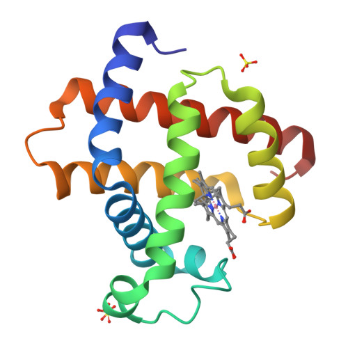
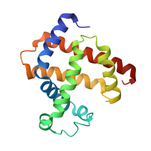
Co(II)/Co(I) reduction-induced axial histidine-flipping in myoglobin reconstituted with a cobalt tetradehydrocorrin as a methionine synthase model.

Hayashi, T.Morita, Y.Mizohata, E.Oohora, K.Ohbayashi, J.Inoue, T.Hisaeda, Y.

(2014) Chem Commun (Camb) 50: 12560-12563

  • DOI: https://doi.org/10.1039/c4cc05448b
  • Primary Citation of Related Structures:  
    3WFT, 3WFU

  • PubMed Abstract: 

    A conjugate between apomyoglobin and cobalt tetradehydrocorrin was prepared to replicate the coordination behavior of cob(I)alamin in methionine synthase. X-ray crystallography reveals that the tetra-coordinated Co(I) species is formed through the cleavage of the axial Co-His93 ligation after the reduction of the penta-coordinated Co(II) cofactor in the heme pocket.


  • Organizational Affiliation

    Department of Applied Chemistry, Graduate School of Engineering, Osaka University, Suita 565-0871, Japan. thayashi@chem.eng.osaka-u.ac.jp.


Macromolecules
Find similar proteins by:  (by identity cutoff)  |  3D Structure
Entity ID: 1
MoleculeChains Sequence LengthOrganismDetailsImage
Myoglobin153Equus caballusMutation(s): 0 
EC: 1.11.1 (UniProt), 1.7 (UniProt)
UniProt
Find proteins for P68082 (Equus caballus)
Explore P68082 
Go to UniProtKB:  P68082
Entity Groups  
Sequence Clusters30% Identity50% Identity70% Identity90% Identity95% Identity100% Identity
UniProt GroupP68082
Sequence Annotations
Expand
  • Reference Sequence
Experimental Data & Validation

Experimental Data

  • Method: X-RAY DIFFRACTION
  • Resolution: 1.35 Å
  • R-Value Free:  0.197 (Depositor), 0.200 (DCC) 
  • R-Value Work:  0.143 (Depositor), 0.140 (DCC) 
  • R-Value Observed: 0.146 (Depositor) 
Space Group: P 1 21 1
Unit Cell:
Length ( Å )Angle ( ˚ )
a = 34.836α = 90
b = 28.72β = 105.6
c = 63.352γ = 90
Software Package:
Software NamePurpose
BSSdata collection
MOLREPphasing
REFMACrefinement
HKL-2000data reduction
HKL-2000data scaling

Structure Validation

View Full Validation Report


Ligand Structure Quality Assessment 

Created with Raphaël 2.3.0Worse 01 BetterLigand structure goodness of fit to experimental dataBest fitted J1SClick on this verticalbar to view detailsBest fitted J1RClick on this verticalbar to view details

Entry History 

Deposition Data

Revision History  (Full details and data files)

  • Version 1.0: 2014-12-03
    Type: Initial release
  • Version 1.1: 2024-03-20
    Changes: Data collection, Database references, Derived calculations