3WRY | pdb_00003wry

Crystal structure of helicase complex 2


Experimental Data Snapshot

  • Method: X-RAY DIFFRACTION
  • Resolution: 2.30 Å
  • R-Value Free: 
    0.248 (Depositor), 0.240 (DCC) 
  • R-Value Work: 
    0.202 (Depositor), 0.190 (DCC) 
  • R-Value Observed: 
    0.202 (Depositor) 

wwPDB Validation   3D Report Full Report


Ligand Structure Quality Assessment 

Created with Raphaël 2.3.0Worse 01 BetterLigand structure goodness of fit to experimental dataBest fitted AGSClick on this verticalbar to view details

This is version 1.3 of the entry. See complete history


Literature

Structural basis for the recognition-evasion arms race between Tomato mosaic virus and the resistance gene Tm-1

Ishibashi, K.Kezuka, Y.Kobayashi, C.Kato, M.Inoue, T.Nonaka, T.Ishikawa, M.Matsumura, H.Katoh, E.

(2014) Proc Natl Acad Sci U S A 111: E3486-E3495

  • DOI: https://doi.org/10.1073/pnas.1407888111
  • Primary Citation of Related Structures:  
    3WRV, 3WRW, 3WRX, 3WRY

  • PubMed Abstract: 

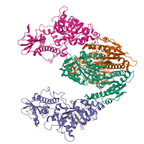
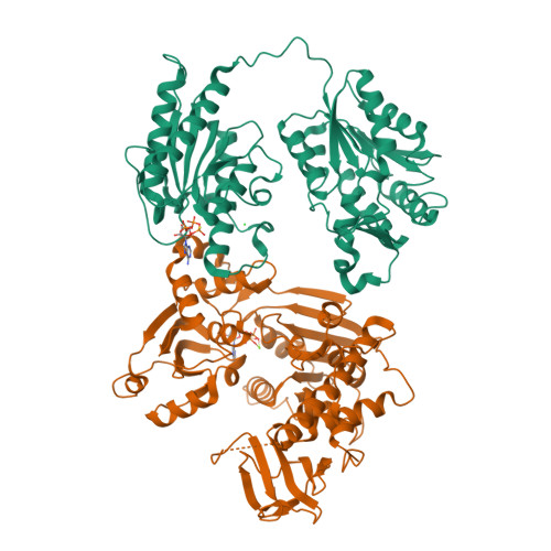
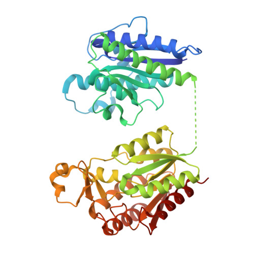
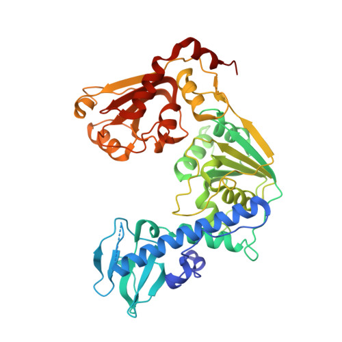
    The tomato mosaic virus (ToMV) resistance gene Tm-1 encodes a protein that shows no sequence homology to functionally characterized proteins. Tm-1 binds ToMV replication proteins and thereby inhibits replication complex formation. ToMV mutants that overcome this resistance have amino acid substitutions in the helicase domain of the replication proteins (ToMV-Hel). A small region of Tm-1 in the genome of the wild tomato Solanum habrochaites has been under positive selection during its antagonistic coevolution with ToMV. Here we report crystal structures for the N-terminal inhibitory domains of Tm-1 and a natural Tm-1 variant with an I91-to-T substitution that has a greater ability to inhibit ToMV RNA replication and their complexes with ToMV-Hel. Each complex contains a Tm-1 dimer and two ToMV-Hel monomers with the interfaces between Tm-1 and ToMV-Hel bridged by ATP. Residues in ToMV-Hel and Tm-1 involved in antagonistic coevolution are found at the interface. The structural differences between ToMV-Hel in its free form and in complex with Tm-1 suggest that Tm-1 affects nucleoside triphosphatase activity of ToMV-Hel, and this effect was confirmed experimentally. Molecular dynamics simulations of complexes formed by Tm-1 with ToMV-Hel variants showed how the amino acid changes in ToMV-Hel impair the interaction with Tm-1 to overcome the resistance. With these findings, together with the biochemical properties of the interactions between ToMV-Hel and Tm-1 variants and effects of the mutations in the polymorphic residues of Tm-1, an atomic view of a step-by-step coevolutionary arms race between a plant resistance protein and a viral protein emerges.


  • Organizational Affiliation

    Plant-Microbe Interactions Research Unit and.


Macromolecules
Find similar proteins by:  (by identity cutoff)  |  3D Structure
Entity ID: 1
MoleculeChains Sequence LengthOrganismDetailsImage
Tm-1 protein
A, B
431Solanum lycopersicumMutation(s): 1 
Gene Names: Tm-1
UniProt
Find proteins for A7M6E7 (Solanum lycopersicum)
Explore A7M6E7 
Go to UniProtKB:  A7M6E7
Entity Groups  
Sequence Clusters30% Identity50% Identity70% Identity90% Identity95% Identity100% Identity
UniProt GroupA7M6E7
Sequence Annotations
Expand
  • Reference Sequence
Find similar proteins by:  (by identity cutoff)  |  3D Structure
Entity ID: 2
MoleculeChains Sequence LengthOrganismDetailsImage
Replicase large subunit
C, D
451Tobacco mosaic virus strain tomato/LMutation(s): 0 
EC: 2.1.1 (PDB Primary Data), 2.7.7 (PDB Primary Data), 2.7.7.48 (PDB Primary Data), 3.6.4.13 (PDB Primary Data)
UniProt
Find proteins for P03587 (Tomato mosaic virus (strain L))
Explore P03587 
Go to UniProtKB:  P03587
Entity Groups  
Sequence Clusters30% Identity50% Identity70% Identity90% Identity95% Identity100% Identity
UniProt GroupP03587
Sequence Annotations
Expand
  • Reference Sequence
Small Molecules
Ligands 3 Unique
IDChains Name / Formula / InChI Key2D Diagram3D Interactions
AGS
Query on AGS

Download Ideal Coordinates CCD File 
E [auth A],
G [auth B],
I [auth C],
K [auth D]
PHOSPHOTHIOPHOSPHORIC ACID-ADENYLATE ESTER
C10 H16 N5 O12 P3 S
NLTUCYMLOPLUHL-KQYNXXCUSA-N
CL
Query on CL

Download Ideal Coordinates CCD File 
H [auth B]CHLORIDE ION
Cl
VEXZGXHMUGYJMC-UHFFFAOYSA-M
MG
Query on MG

Download Ideal Coordinates CCD File 
F [auth A],
J [auth C],
L [auth D],
M [auth D]
MAGNESIUM ION
Mg
JLVVSXFLKOJNIY-UHFFFAOYSA-N
Experimental Data & Validation

Experimental Data

  • Method: X-RAY DIFFRACTION
  • Resolution: 2.30 Å
  • R-Value Free:  0.248 (Depositor), 0.240 (DCC) 
  • R-Value Work:  0.202 (Depositor), 0.190 (DCC) 
  • R-Value Observed: 0.202 (Depositor) 
Space Group: P 21 21 21
Unit Cell:
Length ( Å )Angle ( ˚ )
a = 84.981α = 90
b = 133.55β = 90
c = 195.786γ = 90
Software Package:
Software NamePurpose
HKL-2000data collection
MOLREPphasing
CNSrefinement
HKL-2000data reduction
HKL-2000data scaling

Structure Validation

View Full Validation Report


Ligand Structure Quality Assessment 

Created with Raphaël 2.3.0Worse 01 BetterLigand structure goodness of fit to experimental dataBest fitted AGSClick on this verticalbar to view details

Entry History 

Deposition Data

Revision History  (Full details and data files)

  • Version 1.0: 2014-08-13
    Type: Initial release
  • Version 1.1: 2016-11-16
    Changes: Database references
  • Version 1.2: 2017-11-08
    Changes: Source and taxonomy, Structure summary
  • Version 1.3: 2024-03-20
    Changes: Data collection, Database references, Derived calculations