3ZK1 | pdb_00003zk1

Crystal structure of the sodium binding rotor ring at pH 5.3


Experimental Data Snapshot

  • Method: X-RAY DIFFRACTION
  • Resolution: 2.20 Å
  • R-Value Free: 
    0.234 (Depositor), 0.240 (DCC) 
  • R-Value Work: 
    0.208 (Depositor), 0.210 (DCC) 
  • R-Value Observed: 
    0.209 (Depositor) 

Starting Model: experimental
View more details

wwPDB Validation   3D Report Full Report


Ligand Structure Quality Assessment 

Created with Raphaël 2.3.0Worse 01 BetterLigand structure goodness of fit to experimental dataBest fitted LMTClick on this verticalbar to view detailsBest fitted DMUClick on this verticalbar to view details

This is version 1.2 of the entry. See complete history


Literature

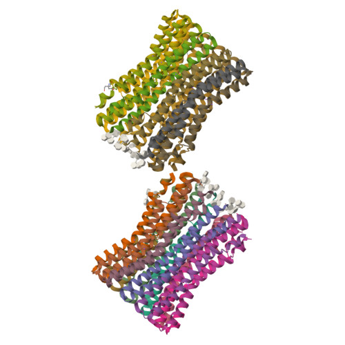
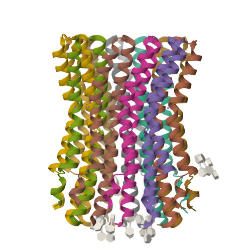
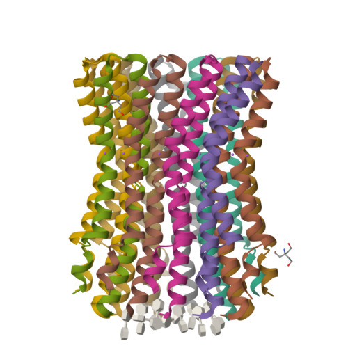
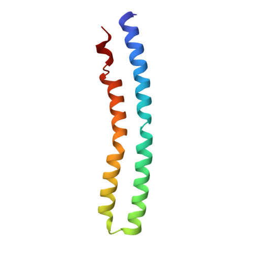
A New Type of Na(+)-Driven ATP Synthase Membrane Rotor with a Two-Carboxylate Ion-Coupling Motif.

Schulz, S.Iglesias-Cans, M.Krah, A.Yildiz, O.Leone, V.Matthies, D.Cook, G.M.Faraldo-Gomez, J.D.Meier, T.

(2013) PLoS Biol 11: 01596

  • DOI: https://doi.org/10.1371/journal.pbio.1001596
  • Primary Citation of Related Structures:  
    3ZK1, 3ZK2

  • PubMed Abstract: 

    The anaerobic bacterium Fusobacterium nucleatum uses glutamate decarboxylation to generate a transmembrane gradient of Na⁺. Here, we demonstrate that this ion-motive force is directly coupled to ATP synthesis, via an F₁F₀-ATP synthase with a novel Na⁺ recognition motif, shared by other human pathogens. Molecular modeling and free-energy simulations of the rotary element of the enzyme, the c-ring, indicate Na⁺ specificity in physiological settings. Consistently, activity measurements showed Na⁺ stimulation of the enzyme, either membrane-embedded or isolated, and ATP synthesis was sensitive to the Na⁺ ionophore monensin. Furthermore, Na⁺ has a protective effect against inhibitors targeting the ion-binding sites, both in the complete ATP synthase and the isolated c-ring. Definitive evidence of Na⁺ coupling is provided by two identical crystal structures of the c₁₁ ring, solved by X-ray crystallography at 2.2 and 2.6 Å resolution, at pH 5.3 and 8.7, respectively. Na⁺ ions occupy all binding sites, each coordinated by four amino acids and a water molecule. Intriguingly, two carboxylates instead of one mediate ion binding. Simulations and experiments demonstrate that this motif implies that a proton is concurrently bound to all sites, although Na⁺ alone drives the rotary mechanism. The structure thus reveals a new mode of ion coupling in ATP synthases and provides a basis for drug-design efforts against this opportunistic pathogen.


  • Organizational Affiliation

    Department of Structural Biology, Max Planck Institute of Biophysics, Frankfurt am Main, Germany.


Macromolecules
Find similar proteins by:  (by identity cutoff)  |  3D Structure
Entity ID: 1
MoleculeChains Sequence LengthOrganismDetailsImage
ATP SYNTHASE SUBUNIT C
A, B, C, D, E
89Fusobacterium nucleatumMutation(s): 0 
Membrane Entity: Yes 
UniProt
Find proteins for Q8RGD7 (Fusobacterium nucleatum subsp. nucleatum (strain ATCC 25586 / DSM 15643 / BCRC 10681 / CIP 101130 / JCM 8532 / KCTC 2640 / LMG 13131 / VPI 4355))
Explore Q8RGD7 
Go to UniProtKB:  Q8RGD7
Entity Groups  
Sequence Clusters30% Identity50% Identity70% Identity90% Identity95% Identity100% Identity
UniProt GroupQ8RGD7
Sequence Annotations
Expand
  • Reference Sequence
Small Molecules
Ligands 4 Unique
IDChains Name / Formula / InChI Key2D Diagram3D Interactions
LMT
Query on LMT

Download Ideal Coordinates CCD File 
AA [auth B],
DA [auth C],
EA [auth C]
DODECYL-BETA-D-MALTOSIDE
C24 H46 O11
NLEBIOOXCVAHBD-QKMCSOCLSA-N
DMU
Query on DMU

Download Ideal Coordinates CCD File 
BB [auth N]
CA [auth C]
DB [auth O]
FB [auth P]
GA [auth D]
DECYL-BETA-D-MALTOPYRANOSIDE
C22 H42 O11
WOQQAWHSKSSAGF-WXFJLFHKSA-N
TAM
Query on TAM

Download Ideal Coordinates CCD File 
KB [auth R],
ZA [auth M]
TRIS(HYDROXYETHYL)AMINOMETHANE
C7 H17 N O3
GKODZWOPPOTFGA-UHFFFAOYSA-N
NA
Query on NA

Download Ideal Coordinates CCD File 
AB [auth N]
BA [auth C]
CB [auth O]
EB [auth P]
FA [auth D]
SODIUM ION
Na
FKNQFGJONOIPTF-UHFFFAOYSA-N
Experimental Data & Validation

Experimental Data

  • Method: X-RAY DIFFRACTION
  • Resolution: 2.20 Å
  • R-Value Free:  0.234 (Depositor), 0.240 (DCC) 
  • R-Value Work:  0.208 (Depositor), 0.210 (DCC) 
  • R-Value Observed: 0.209 (Depositor) 
Space Group: P 1 21 1
Unit Cell:
Length ( Å )Angle ( ˚ )
a = 135.77α = 90
b = 83.9β = 112.85
c = 150.99γ = 90
Software Package:
Software NamePurpose
PHENIXrefinement
XDSdata reduction
XSCALEdata scaling
PHASERphasing

Structure Validation

View Full Validation Report


Ligand Structure Quality Assessment 

Created with Raphaël 2.3.0Worse 01 BetterLigand structure goodness of fit to experimental dataBest fitted LMTClick on this verticalbar to view detailsBest fitted DMUClick on this verticalbar to view details

Entry History 

Deposition Data

Revision History  (Full details and data files)

  • Version 1.0: 2013-05-29
    Type: Initial release
  • Version 1.1: 2013-09-18
    Changes: Database references, Other
  • Version 1.2: 2023-12-20
    Changes: Data collection, Database references, Derived calculations, Other, Refinement description