4ZM4 | pdb_00004zm4

Complex structure of PctV K276R mutant with PMP and 3-dehydroshkimate


Experimental Data Snapshot

  • Method: X-RAY DIFFRACTION
  • Resolution: 2.40 Å
  • R-Value Free: 
    0.278 (Depositor), 0.280 (DCC) 
  • R-Value Work: 
    0.238 (Depositor), 0.250 (DCC) 
  • R-Value Observed: 
    0.240 (Depositor) 

Starting Model: experimental
View more details

wwPDB Validation   3D Report Full Report


Ligand Structure Quality Assessment 

Created with Raphaël 2.3.0Worse 01 BetterLigand structure goodness of fit to experimental dataBest fitted P3BClick on this verticalbar to view detailsBest fitted PLPClick on this verticalbar to view details

This is version 1.3 of the entry. See complete history


Literature

Mechanism-Based Trapping of the Quinonoid Intermediate by Using the K276R Mutant of PLP-Dependent 3-Aminobenzoate Synthase PctV in the Biosynthesis of Pactamycin.

Hirayama, A.Miyanaga, A.Kudo, F.Eguchi, T.

(2015) Chembiochem 16: 2484-2490

  • DOI: https://doi.org/10.1002/cbic.201500426
  • Primary Citation of Related Structures:  
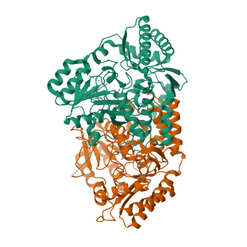
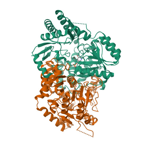
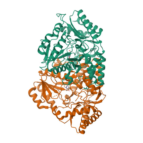
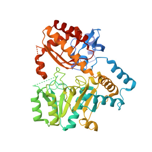
    4ZM3, 4ZM4

  • PubMed Abstract: 

    Mutational analysis of the pyridoxal 5'-phosphate (PLP)-dependent enzyme PctV was carried out to elucidate the multi-step reaction mechanism for the formation of 3-aminobenzoate (3-ABA) from 3-dehydroshikimate (3-DSA). Introduction of mutation K276R led to the accumulation of a quinonoid intermediate with an absorption maximum at 580 nm after the reaction of pyridoxamine 5'-phosphate (PMP) with 3-DSA. The chemical structure of this intermediate was supported by X-ray crystallographic analysis of the complex formed between the K276R mutant and the quinonoid intermediate. These results clearly show that a quinonoid intermediate is involved in the formation of 3-ABA. They also indicate that Lys276 (in the active site of PctV) plays multiple roles, including acid/base catalysis during the dehydration reaction of the quinonoid intermediate.


  • Organizational Affiliation

    Department of Chemistry and Materials Science, Tokyo Institute of Technology, 2-12-1 O-okayama, Meguro-ku, Tokyo, 152-8551, Japan.


Macromolecules
Find similar proteins by:  (by identity cutoff)  |  3D Structure
Entity ID: 1
MoleculeChains Sequence LengthOrganismDetailsImage
Aminotransferase
A, B, C, D, E
447Streptomyces pactumMutation(s): 1 
Gene Names: pctVptmT
UniProt
Find proteins for A8R0K5 (Streptomyces pactum)
Explore A8R0K5 
Go to UniProtKB:  A8R0K5
Entity Groups  
Sequence Clusters30% Identity50% Identity70% Identity90% Identity95% Identity100% Identity
UniProt GroupA8R0K5
Sequence Annotations
Expand
  • Reference Sequence
Experimental Data & Validation

Experimental Data

  • Method: X-RAY DIFFRACTION
  • Resolution: 2.40 Å
  • R-Value Free:  0.278 (Depositor), 0.280 (DCC) 
  • R-Value Work:  0.238 (Depositor), 0.250 (DCC) 
  • R-Value Observed: 0.240 (Depositor) 
Space Group: H 3 2
Unit Cell:
Length ( Å )Angle ( ˚ )
a = 178.93α = 90
b = 178.93β = 90
c = 467.899γ = 120
Software Package:
Software NamePurpose
REFMACrefinement
MOSFLMdata reduction
MOSFLMdata scaling
MOLREPphasing
Cootmodel building

Structure Validation

View Full Validation Report


Ligand Structure Quality Assessment 

Created with Raphaël 2.3.0Worse 01 BetterLigand structure goodness of fit to experimental dataBest fitted P3BClick on this verticalbar to view detailsBest fitted PLPClick on this verticalbar to view details

Entry History & Funding Information

Deposition Data


Funding OrganizationLocationGrant Number
Ministry of Education, Culture, Sports, Science and TechnologyJapan22108003

Revision History  (Full details and data files)

  • Version 1.0: 2015-10-14
    Type: Initial release
  • Version 1.1: 2016-01-27
    Changes: Database references
  • Version 1.2: 2020-02-19
    Changes: Data collection, Derived calculations
  • Version 1.3: 2023-11-08
    Changes: Data collection, Database references, Refinement description