1BDG | pdb_00001bdg

HEXOKINASE FROM SCHISTOSOMA MANSONI COMPLEXED WITH GLUCOSE


Experimental Data Snapshot

  • Method: X-RAY DIFFRACTION
  • Resolution: 2.60 Å
  • R-Value Free: 
    0.256 (Depositor) 
  • R-Value Work: 
    0.173 (Depositor), 0.190 (DCC) 
  • R-Value Observed: 
    0.173 (Depositor) 

Starting Model: experimental
View more details

wwPDB Validation   3D Report Full Report


Ligand Structure Quality Assessment 

Created with Raphaël 2.3.0Worse 01 BetterLigand structure goodness of fit to experimental dataBest fitted GLCClick on this verticalbar to view details

This is version 1.6 of the entry. See complete history


Literature

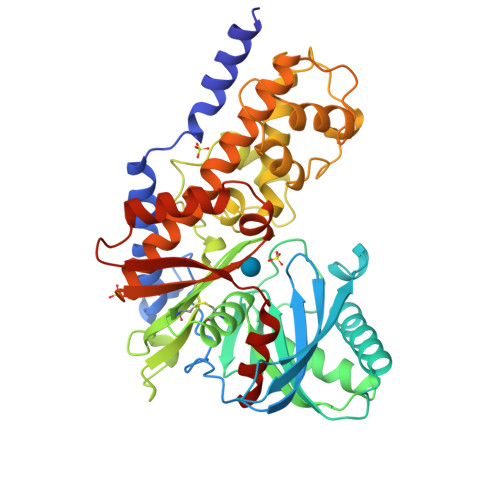
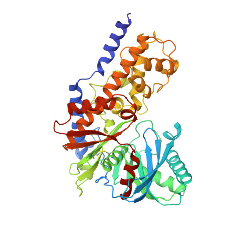
The structure of mammalian hexokinase-1.

Mulichak, A.M.Wilson, J.E.Padmanabhan, K.Garavito, R.M.

(1998) Nat Struct Biol 5: 555-560

  • DOI: https://doi.org/10.1038/811
  • Primary Citation of Related Structures:  
    1BDG, 1BG3

  • PubMed Abstract: 

    We have determined the structures of the glucose-6-phosphate (G6P)-inhibitable 100,000 Mr Type I hexokinase from rat and the G6P-sensitive 50,000 Mr hexokinase from Schistosoma mansoni at a resolution of 2.8 and 2.6 A respectively. The structures define the glucose and G6P binding sites in these enzymes, suggest the mechanisms of intradomain G6P inhibition and activity loss in the Type I hexokinase N-terminal half, and reveal the structure of the membrane targeting motif that integrates the Type I hexokinase into the outer mitochondrial membrane.


  • Organizational Affiliation

    Department of Biochemistry, Michigan State University, East Lansing, USA. mulichak@alecto.bch.msu.edu


Macromolecules
Find similar proteins by:  (by identity cutoff)  |  3D Structure
Entity ID: 1
MoleculeChains Sequence LengthOrganismDetailsImage
HEXOKINASE451Schistosoma mansoniMutation(s): 0 
EC: 2.7.1.1
UniProt
Find proteins for Q26609 (Schistosoma mansoni)
Explore Q26609 
Go to UniProtKB:  Q26609
Entity Groups  
Sequence Clusters30% Identity50% Identity70% Identity90% Identity95% Identity100% Identity
UniProt GroupQ26609
Sequence Annotations
Expand
  • Reference Sequence
Experimental Data & Validation

Experimental Data

  • Method: X-RAY DIFFRACTION
  • Resolution: 2.60 Å
  • R-Value Free:  0.256 (Depositor) 
  • R-Value Work:  0.173 (Depositor), 0.190 (DCC) 
  • R-Value Observed: 0.173 (Depositor) 
Space Group: P 61 2 2
Unit Cell:
Length ( Å )Angle ( ˚ )
a = 114.3α = 90
b = 114.3β = 90
c = 155.2γ = 120
Software Package:
Software NamePurpose
R-AXISdata collection
R-AXISdata reduction
AMoREphasing
X-PLORrefinement
R-AXISdata scaling

Structure Validation

View Full Validation Report


Ligand Structure Quality Assessment 

Created with Raphaël 2.3.0Worse 01 BetterLigand structure goodness of fit to experimental dataBest fitted GLCClick on this verticalbar to view details

Entry History 

Deposition Data

Revision History  (Full details and data files)

  • Version 1.0: 1999-05-11
    Type: Initial release
  • Version 1.1: 2008-03-24
    Changes: Version format compliance
  • Version 1.2: 2011-07-13
    Changes: Version format compliance
  • Version 1.3: 2020-07-29
    Type: Remediation
    Reason: Carbohydrate remediation
    Changes: Data collection, Derived calculations, Structure summary
  • Version 1.4: 2022-12-21
    Changes: Database references, Structure summary
  • Version 1.5: 2023-08-09
    Changes: Refinement description
  • Version 1.6: 2024-10-16
    Changes: Data collection, Structure summary