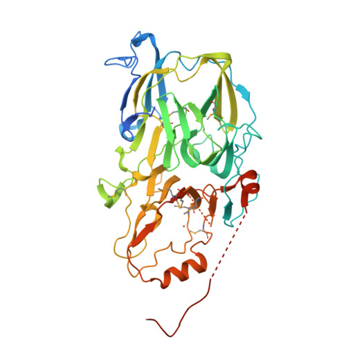
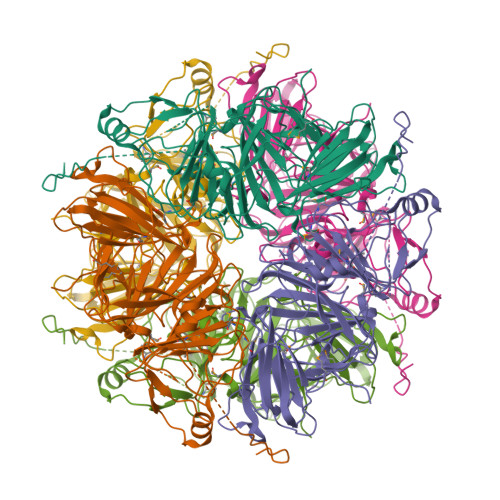
1EY2

HUMAN HOMOGENTISATE DIOXYGENASE WITH FE(II)


Experimental Data Snapshot

  • Method: X-RAY DIFFRACTION
  • Resolution: 2.30 Å
  • R-Value Free: 0.242 
  • R-Value Work: 0.193 
  • R-Value Observed: 0.193 

wwPDB Validation   3D Report Full Report


This is version 1.4 of the entry. See complete history


Literature

Crystal structure of human homogentisate dioxygenase.

Titus, G.P.Mueller, H.A.Burgner, J.Rodriguez De Cordoba, S.Penalva, M.A.Timm, D.E.

(2000) Nat Struct Biol 7: 542-546

  • DOI: https://doi.org/10.1038/76756
  • Primary Citation of Related Structures:  
    1EY2, 1EYB

  • PubMed Abstract: 

    Homogentisate dioxygenase (HGO) cleaves the aromatic ring during the metabolic degradation of Phe and Tyr. HGO deficiency causes alkaptonuria (AKU), the first human disease shown to be inherited as a recessive Mendelian trait. Crystal structures of apo-HGO and HGO containing an iron ion have been determined at 1.9 and 2.3 A resolution, respectively. The HGO protomer, which contains a 280-residue N-terminal domain and a 140-residue C-terminal domain, associates as a hexamer arranged as a dimer of trimers. The active site iron ion is coordinated near the interface between subunits in the HGO trimer by a Glu and two His side chains. HGO represents a new structural class of dioxygenases. The largest group of AKU associated missense mutations affect residues located in regions of contact between subunits.


  • Organizational Affiliation

    Department of Biochemistry and Molecular Biology, Indiana University School of Medicine, 635 Barnhill Drive, Indianapolis, Indiana 46202, USA.


Macromolecules
Find similar proteins by:  (by identity cutoff)  |  3D Structure
Entity ID: 1
MoleculeChains Sequence LengthOrganismDetailsImage
HOMOGENTISATE 1,2-DIOXYGENASE471Homo sapiensMutation(s): 10 
EC: 1.13.11.5
UniProt & NIH Common Fund Data Resources
Find proteins for Q93099 (Homo sapiens)
Explore Q93099 
Go to UniProtKB:  Q93099
PHAROS:  Q93099
GTEx:  ENSG00000113924 
Entity Groups  
Sequence Clusters30% Identity50% Identity70% Identity90% Identity95% Identity100% Identity
UniProt GroupQ93099
Sequence Annotations
Expand
  • Reference Sequence
Small Molecules
Ligands 1 Unique
IDChains Name / Formula / InChI Key2D Diagram3D Interactions
FE2
Query on FE2

Download Ideal Coordinates CCD File 
B [auth A]FE (II) ION
Fe
CWYNVVGOOAEACU-UHFFFAOYSA-N
Modified Residues  1 Unique
IDChains TypeFormula2D DiagramParent
MSE
Query on MSE
A
L-PEPTIDE LINKINGC5 H11 N O2 SeMET
Experimental Data & Validation

Experimental Data

  • Method: X-RAY DIFFRACTION
  • Resolution: 2.30 Å
  • R-Value Free: 0.242 
  • R-Value Work: 0.193 
  • R-Value Observed: 0.193 
  • Space Group: P 63 2 2
Unit Cell:
Length ( Å )Angle ( ˚ )
a = 157.809α = 90
b = 157.809β = 90
c = 95.147γ = 120
Software Package:
Software NamePurpose
MLPHAREphasing
X-PLORrefinement
DENZOdata reduction
SCALEPACKdata scaling

Structure Validation

View Full Validation Report


Entry History 

Deposition Data

Revision History  (Full details and data files)

  • Version 1.0: 2000-11-05
    Type: Initial release
  • Version 1.1: 2008-04-27
    Changes: Version format compliance
  • Version 1.2: 2011-07-13
    Changes: Derived calculations, Version format compliance
  • Version 1.3: 2021-11-03
    Changes: Database references, Derived calculations
  • Version 1.4: 2024-11-20
    Changes: Data collection, Structure summary