1I6M | pdb_00001i6m

1.7 HIGH RESOLUTION EXPERIMENTAL PHASES FOR TRYPTOPHANYL-TRNA SYNTHETASE COMPLEXED WITH TRYPTOPHANYL-5'AMP


Experimental Data Snapshot

  • Method: X-RAY DIFFRACTION
  • Resolution: 1.72 Å
  • R-Value Free: 
    0.257 (Depositor), 0.210 (DCC) 
  • R-Value Work: 
    0.225 (Depositor), 0.210 (DCC) 

wwPDB Validation   3D Report Full Report


Ligand Structure Quality Assessment 

Created with Raphaël 2.3.0Worse 01 BetterLigand structure goodness of fit to experimental dataBest fitted TYMClick on this verticalbar to view details

This is version 1.3 of the entry. See complete history


Literature

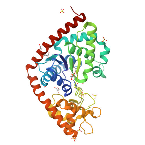
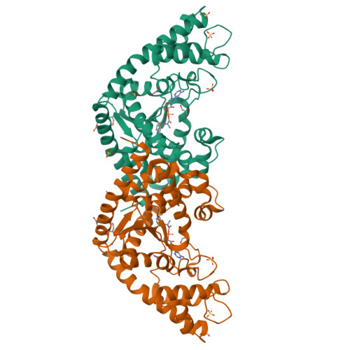
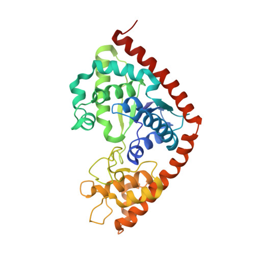
High-resolution experimental phases for tryptophanyl-tRNA synthetase (TrpRS) complexed with tryptophanyl-5'AMP.

Retailleau, P.Yin, Y.Hu, M.Roach, J.Bricogne, G.Vonrhein, C.Roversi, P.Blanc, E.Sweet, R.M.Carter, C.W.

(2001) Acta Crystallogr D Biol Crystallogr 57: 1595-1608

  • DOI: https://doi.org/10.1107/s090744490101215x
  • Primary Citation of Related Structures:  
    1I6K, 1I6L, 1I6M

  • PubMed Abstract: 

    Native data, anomalous data at three wavelengths and an independent peak-wavelength data set for SeMet-substituted protein have been collected from cryoprotected crystals of the TrpRS-adenylate product (TAM) complex to a resolution limit of 1.7 A. Independent phase sets were developed using SHARP and improved by solvent flipping with SOLOMON using molecular envelopes derived from experimental densities for, respectively, peak-wavelength SAD data from four different crystals, MAD data and their M(S)IRAS combinations with native data. Hendrickson-Lattman phase-probability coefficients from each phase set were used in BUSTER to drive maximum-likelihood refinements of well defined parts of the previously refined room-temperature 2.9 A structure. Maximum-entropy completion followed by manual rebuilding was then used to generate a model for the missing segments, bound ligand and solvent molecules. Surprisingly, peak-wavelength SAD experiments produced the smallest phase errors relative to the refined structures. Selenomethionylated models deviate from one another by 0.25 A and from the native model by 0.38 A, but all have r.m.s. deviations of approximately 1.0 A from the 2.9 A model. Difference Fourier calculations between amplitudes from the 300 K experiment and the new amplitudes at 100 K using 1.7 A model phases show no significant structural changes arising from temperature variation or addition of cryoprotectant. The main differences between low- and high-resolution structures arise from correcting side-chain rotamers in the core of the protein as well as on the surface. These changes improve various structure-validation criteria.


  • Organizational Affiliation

    Department of Biochemistry and Biophysics, CB# 7260, University of North Carolina at Chapel Hill, Chapel Hill, NC, USA.


Macromolecules
Find similar proteins by:  (by identity cutoff)  |  3D Structure
Entity ID: 1
MoleculeChains Sequence LengthOrganismDetailsImage
TRYPTOPHANYL-TRNA SYNTHETASE328Geobacillus stearothermophilusMutation(s): 0 
EC: 6.1.1.2
UniProt
Find proteins for P00953 (Geobacillus stearothermophilus)
Explore P00953 
Go to UniProtKB:  P00953
Entity Groups  
Sequence Clusters30% Identity50% Identity70% Identity90% Identity95% Identity100% Identity
UniProt GroupP00953
Sequence Annotations
Expand
  • Reference Sequence
Small Molecules
Ligands 4 Unique
IDChains Name / Formula / InChI Key2D Diagram3D Interactions
TYM
Query on TYM

Download Ideal Coordinates CCD File 
J [auth A]TRYPTOPHANYL-5'AMP
C21 H24 N7 O8 P
IFQVDHDRFCKAAW-SQIXAUHQSA-N
SO4
Query on SO4

Download Ideal Coordinates CCD File 
B [auth A]
C [auth A]
D [auth A]
E [auth A]
F [auth A]
SULFATE ION
O4 S
QAOWNCQODCNURD-UHFFFAOYSA-L
GOL
Query on GOL

Download Ideal Coordinates CCD File 
K [auth A],
L [auth A],
M [auth A],
N [auth A]
GLYCEROL
C3 H8 O3
PEDCQBHIVMGVHV-UHFFFAOYSA-N
NH4
Query on NH4

Download Ideal Coordinates CCD File 
I [auth A]AMMONIUM ION
H4 N
QGZKDVFQNNGYKY-UHFFFAOYSA-O
Experimental Data & Validation

Experimental Data

  • Method: X-RAY DIFFRACTION
  • Resolution: 1.72 Å
  • R-Value Free:  0.257 (Depositor), 0.210 (DCC) 
  • R-Value Work:  0.225 (Depositor), 0.210 (DCC) 
Space Group: P 43 21 2
Unit Cell:
Length ( Å )Angle ( ˚ )
a = 59.77α = 90
b = 59.77β = 90
c = 232.65γ = 90
Software Package:
Software NamePurpose
SHARPphasing
BUSTER-TNTrefinement
DENZOdata reduction
SCALEPACKdata scaling
BUSTER-TNTphasing

Structure Validation

View Full Validation Report


Ligand Structure Quality Assessment 

Created with Raphaël 2.3.0Worse 01 BetterLigand structure goodness of fit to experimental dataBest fitted TYMClick on this verticalbar to view details

Entry History 

Deposition Data

Revision History  (Full details and data files)

  • Version 1.0: 2001-11-07
    Type: Initial release
  • Version 1.1: 2008-04-27
    Changes: Version format compliance
  • Version 1.2: 2011-07-13
    Changes: Derived calculations, Version format compliance
  • Version 1.3: 2024-02-07
    Changes: Data collection, Database references, Derived calculations