1OEL | pdb_00001oel

CONFORMATIONAL VARIABILITY IN THE REFINED STRUCTURE OF THE CHAPERONIN GROEL AT 2.8 ANGSTROM RESOLUTION


Experimental Data Snapshot

  • Method: X-RAY DIFFRACTION
  • Resolution: 2.80 Å
  • R-Value Free: 
    0.270 (Depositor) 
  • R-Value Work: 
    0.227 (Depositor) 
  • R-Value Observed: 
    0.227 (Depositor) 

wwPDB Validation   3D Report Full Report


This is version 1.4 of the entry. See complete history


Literature

Conformational variability in the refined structure of the chaperonin GroEL at 2.8 A resolution.

Braig, K.Adams, P.D.Brunger, A.T.

(1995) Nat Struct Biol 2: 1083-1094

  • DOI: https://doi.org/10.1038/nsb1295-1083
  • Primary Citation of Related Structures:  
    1OEL

  • PubMed Abstract: 

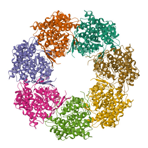
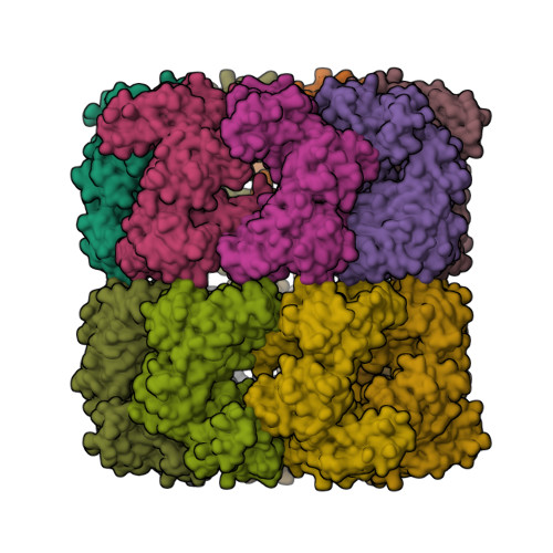
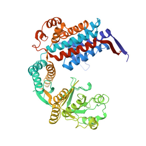
    Improved refinement of the crystal structure of GroEL from Escherichia coli has resulted in a complete atomic model for the first 524 residues. A new torsion-angle dynamics method and non-crystallographic symmetry restraints were used in the refinement. The model indicates that conformational variability exists due to rigid-body movements between the apical and intermediate domains of GroEL, resulting in deviations from strict seven-fold symmetry. The regions of the protein involved in polypeptide and GroES binding show unusually high B factors; these values may indicate mobility or discrete disorder. The variability of these regions may play a role in the ability of GroEL to bind a wide variety of substrates.


  • Organizational Affiliation

    Department of Genetics, Yale University, New Haven, Connecticut 06510, USA.


Macromolecules
Find similar proteins by:  (by identity cutoff)  |  3D Structure
Entity ID: 1
MoleculeChains Sequence LengthOrganismDetailsImage
GROEL (HSP60 CLASS)
A, B, C, D, E
547Escherichia coliMutation(s): 2 
Gene Names: GROEL
EC: 5.6.1.7
UniProt
Find proteins for P0A6F5 (Escherichia coli (strain K12))
Explore P0A6F5 
Go to UniProtKB:  P0A6F5
Entity Groups  
Sequence Clusters30% Identity50% Identity70% Identity90% Identity95% Identity100% Identity
UniProt GroupP0A6F5
Sequence Annotations
Expand
  • Reference Sequence
Experimental Data & Validation

Experimental Data

  • Method: X-RAY DIFFRACTION
  • Resolution: 2.80 Å
  • R-Value Free:  0.270 (Depositor) 
  • R-Value Work:  0.227 (Depositor) 
  • R-Value Observed: 0.227 (Depositor) 
Space Group: C 2 2 21
Unit Cell:
Length ( Å )Angle ( ˚ )
a = 178.38α = 90
b = 204.98β = 90
c = 280.98γ = 90
Software Package:
Software NamePurpose
X-PLORmodel building
X-PLORrefinement
X-PLORphasing

Structure Validation

View Full Validation Report


Entry History 

Deposition Data

Revision History  (Full details and data files)

  • Version 1.0: 1996-04-03
    Type: Initial release
  • Version 1.1: 2008-03-24
    Changes: Version format compliance
  • Version 1.2: 2011-07-13
    Changes: Version format compliance
  • Version 1.3: 2021-11-03
    Changes: Database references, Other
  • Version 1.4: 2024-02-14
    Changes: Data collection