1QKO | pdb_00001qko

HIGH RESOLUTION X-RAY STRUCTURE OF AN EARLY INTERMEDIATE IN THE BACTERIORHODOPSIN PHOTOCYCLE


Experimental Data Snapshot

  • Method: X-RAY DIFFRACTION
  • Resolution: 2.10 Å
  • R-Value Free: 
    0.303 (Depositor), 0.240 (DCC) 
  • R-Value Work: 
    0.261 (Depositor), 0.220 (DCC) 
  • R-Value Observed: 
    0.261 (Depositor) 

Starting Model: experimental
View more details

wwPDB Validation   3D Report Full Report


Ligand Structure Quality Assessment 

Created with Raphaël 2.3.0Worse 01 BetterLigand structure goodness of fit to experimental dataBest fitted RETClick on this verticalbar to view details

This is version 1.5 of the entry. See complete history


Literature

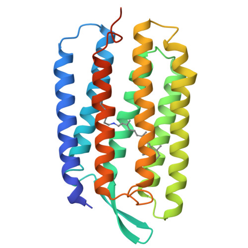
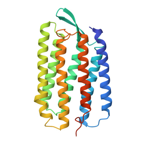
High Resolution X-Ray Structure of an Early Intermediate in the Bacteriorhodopsin Photocycle

Edman, K.Nollert, P.Royant, A.Belrhali, H.Pebay-Peyroula, E.Hajdu, J.Neutze, R.Landau, E.M.

(1999) Nature 401: 822

  • DOI: https://doi.org/10.1038/44623
  • Primary Citation of Related Structures:  
    1QKO, 1QKP

  • PubMed Abstract: 

    Bacteriorhodopsin is the simplest known photon-driven proton pump and as such provides a model for the study of a basic function in bioenergetics. Its seven transmembrane helices encompass a proton translocation pathway containing the chromophore, a retinal molecule covalently bound to lysine 216 through a protonated Schiff base, and a series of proton donors and acceptors. Photoisomerization of the all-trans retinal to the 13-cis configuration initiates the vectorial translocation of a proton from the Schiff base, the primary proton donor, to the extracellular side, followed by reprotonation of the Schiff base from the cytoplasm. Here we describe the high-resolution X-ray structure of an early intermediate in the photocycle of bacteriorhodopsin, which is formed directly after photoexcitation. A key water molecule is dislocated, allowing the primary proton acceptor, Asp 85, to move. Movement of the main-chain Lys 216 locally disrupts the hydrogen-bonding network of helix G, facilitating structural changes later in the photocycle.


  • Organizational Affiliation

    Department of Biochemistry, Uppsala University, Biomedical Centre, Sweden.


Macromolecules
Find similar proteins by:  (by identity cutoff)  |  3D Structure
Entity ID: 1
MoleculeChains Sequence LengthOrganismDetailsImage
BACTERIORHODOPSIN248Halobacterium salinarumMutation(s): 0 
Membrane Entity: Yes 
UniProt
Find proteins for P02945 (Halobacterium salinarum (strain ATCC 700922 / JCM 11081 / NRC-1))
Explore P02945 
Go to UniProtKB:  P02945
Entity Groups  
Sequence Clusters30% Identity50% Identity70% Identity90% Identity95% Identity100% Identity
UniProt GroupP02945
Sequence Annotations
Expand
  • Reference Sequence
Small Molecules
Ligands 1 Unique
IDChains Name / Formula / InChI Key2D Diagram3D Interactions
RET
Query on RET

Download Ideal Coordinates CCD File 
B [auth A]RETINAL
C20 H28 O
NCYCYZXNIZJOKI-OVSJKPMPSA-N
Experimental Data & Validation

Experimental Data

  • Method: X-RAY DIFFRACTION
  • Resolution: 2.10 Å
  • R-Value Free:  0.303 (Depositor), 0.240 (DCC) 
  • R-Value Work:  0.261 (Depositor), 0.220 (DCC) 
  • R-Value Observed: 0.261 (Depositor) 
Space Group: P 63
Unit Cell:
Length ( Å )Angle ( ˚ )
a = 61.06α = 90
b = 61.06β = 90
c = 110.57γ = 120
Software Package:
Software NamePurpose
CNSrefinement
DENZOdata reduction
CCP4data scaling
CNSphasing

Structure Validation

View Full Validation Report


Ligand Structure Quality Assessment 

Created with Raphaël 2.3.0Worse 01 BetterLigand structure goodness of fit to experimental dataBest fitted RETClick on this verticalbar to view details

Entry History 

Deposition Data

Revision History  (Full details and data files)

  • Version 1.0: 1999-10-24
    Type: Initial release
  • Version 1.1: 2011-05-08
    Changes: Version format compliance
  • Version 1.2: 2011-07-13
    Changes: Version format compliance
  • Version 1.3: 2019-05-15
    Changes: Data collection, Derived calculations, Experimental preparation, Other
  • Version 1.4: 2023-12-13
    Changes: Data collection, Database references, Derived calculations, Other, Refinement description
  • Version 1.5: 2024-11-13
    Changes: Structure summary