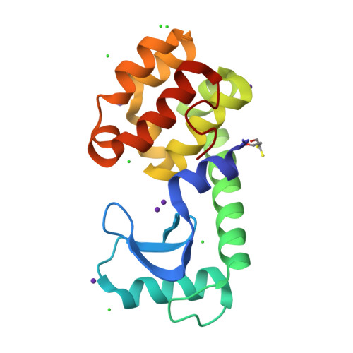
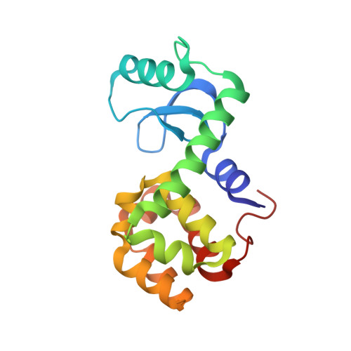
1SX2

Use of a Halide Binding Site to Bypass the 1000-atom Limit to Structure Determination by Direct Methods


Experimental Data Snapshot

  • Method: X-RAY DIFFRACTION
  • Resolution: 1.06 Å
  • R-Value Free: 0.143 
  • R-Value Work: 0.125 
  • R-Value Observed: 0.121 

wwPDB Validation   3D Report Full Report


This is version 1.6 of the entry. See complete history


Literature

Use of an ion-binding site to bypass the 1000-atom limit to structure determination by direct methods.

Mooers, B.H.Matthews, B.W.

(2004) Acta Crystallogr D Biol Crystallogr 60: 1726-1737

  • DOI: https://doi.org/10.1107/S0907444904017020
  • Primary Citation of Related Structures:  
    1SWY, 1SWZ, 1SX2, 1SX7

  • PubMed Abstract: 

    Proteins with more than 1000 non-H atoms and without heavy-atom prosthetic groups are very difficult to solve by ab initio direct methods. T4 lysozyme is being used to explore these limits. The protein has 1309 non-H atoms, seven S atoms, no disulfide bonds and no heavy-atom prosthetic group. It is recalcitrant to structure determination by direct methods using X-ray diffraction data to 0.97 A. It is shown here that it is possible to obtain a truly ab initio structure determination of a variant of the protein that has an Rb+ (Z = 37) binding site. Using diffraction data to 1.06 A resolution, the direct-methods programs SIR2002 and ACORN independently solved the structure in about 20 h. The bound Rb+, which contributes about 1.7% of the total scattering, does not appear to distort the structure or to inhibit refinement (R factor 12.1%). The phases obtained via SIR2002 or ACORN are in good agreement with those from a reference structure obtained from conventional molecular-substitution and refinement procedures (average error in the figure-of-merit-weighted phases of less than 25 degrees). Thus, proteins with more than 1000 atoms that include halide-binding or other such sites may be amenable to structure determination by ab initio direct methods. The direct-methods approaches are also compared with structure determination via use of the anomalous scattering of the Rb+ ion. As shown by examples, high-resolution structures determined by direct methods can be useful in highlighting regions of strain in the protein, including short hydrogen bonds and non-planar peptide groups.


  • Organizational Affiliation

    Institute of Molecular Biology, Howard Hughes Medical Institute and Department of Physics, 1229 University of Oregon, Eugene, OR 97403-1229, USA.


Macromolecules
Find similar proteins by:  (by identity cutoff)  |  3D Structure
Entity ID: 1
MoleculeChains Sequence LengthOrganismDetailsImage
Lysozyme164Tequatrovirus T4Mutation(s): 2 
Gene Names: E
EC: 3.2.1.17
UniProt
Find proteins for P00720 (Enterobacteria phage T4)
Explore P00720 
Go to UniProtKB:  P00720
Entity Groups  
Sequence Clusters30% Identity50% Identity70% Identity90% Identity95% Identity100% Identity
UniProt GroupP00720
Sequence Annotations
Expand
  • Reference Sequence
Small Molecules
Ligands 3 Unique
IDChains Name / Formula / InChI Key2D Diagram3D Interactions
RB
Query on RB

Download Ideal Coordinates CCD File 
B [auth A],
C [auth A],
D [auth A],
E [auth A],
F [auth A]
RUBIDIUM ION
Rb
NCCSSGKUIKYAJD-UHFFFAOYSA-N
BME
Query on BME

Download Ideal Coordinates CCD File 
L [auth A],
M [auth A]
BETA-MERCAPTOETHANOL
C2 H6 O S
DGVVWUTYPXICAM-UHFFFAOYSA-N
CL
Query on CL

Download Ideal Coordinates CCD File 
G [auth A],
H [auth A],
I [auth A],
J [auth A],
K [auth A]
CHLORIDE ION
Cl
VEXZGXHMUGYJMC-UHFFFAOYSA-M
Experimental Data & Validation

Experimental Data

  • Method: X-RAY DIFFRACTION
  • Resolution: 1.06 Å
  • R-Value Free: 0.143 
  • R-Value Work: 0.125 
  • R-Value Observed: 0.121 
  • Space Group: P 32 2 1
Unit Cell:
Length ( Å )Angle ( ˚ )
a = 60.199α = 90
b = 60.199β = 90
c = 95.242γ = 120
Software Package:
Software NamePurpose
MAR345data collection
SCALAdata scaling
ACORNphasing
SHELXL-97refinement
MOSFLMdata reduction
CCP4data scaling

Structure Validation

View Full Validation Report


Entry History 

Deposition Data

Revision History  (Full details and data files)

  • Version 1.0: 2004-11-23
    Type: Initial release
  • Version 1.1: 2008-04-30
    Changes: Version format compliance
  • Version 1.2: 2011-07-13
    Changes: Version format compliance
  • Version 1.3: 2017-10-11
    Changes: Refinement description
  • Version 1.4: 2018-01-31
    Changes: Experimental preparation
  • Version 1.5: 2021-10-27
    Changes: Database references, Derived calculations
  • Version 1.6: 2024-02-14
    Changes: Data collection