1UKC | pdb_00001ukc

Crystal Structure of Aspergillus niger EstA


Experimental Data Snapshot

  • Method: X-RAY DIFFRACTION
  • Resolution: 2.10 Å
  • R-Value Free: 
    0.184 (Depositor), 0.190 (DCC) 
  • R-Value Work: 
    0.158 (Depositor), 0.170 (DCC) 
  • R-Value Observed: 
    0.158 (Depositor) 

Starting Models: experimental
View more details

wwPDB Validation   3D Report Full Report


Ligand Structure Quality Assessment 

Created with Raphaël 2.3.0Worse 01 BetterLigand structure goodness of fit to experimental dataBest fitted NAGClick on this verticalbar to view detailsBest fitted MANClick on this verticalbar to view details

This is version 2.2 of the entry. See complete history


Literature

Aspergillus niger protein EstA defines a new class of fungal esterases within the alpha/beta hydrolase fold superfamily of proteins

Bourne, Y.Hasper, A.A.Chahinian, H.Juin, M.De Graaff, L.H.Marchot, P.

(2004) Structure 12: 677-687

  • DOI: https://doi.org/10.1016/j.str.2004.03.005
  • Primary Citation of Related Structures:  
    1UKC

  • PubMed Abstract: 

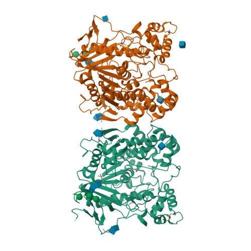
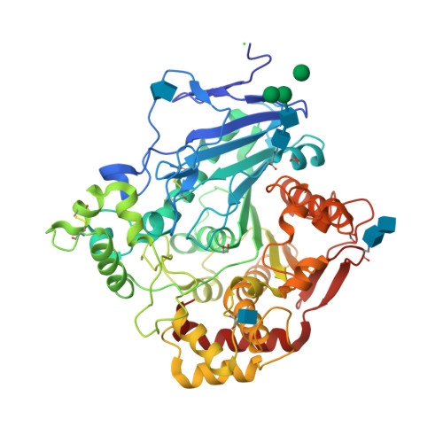
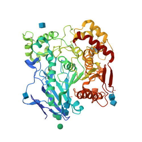
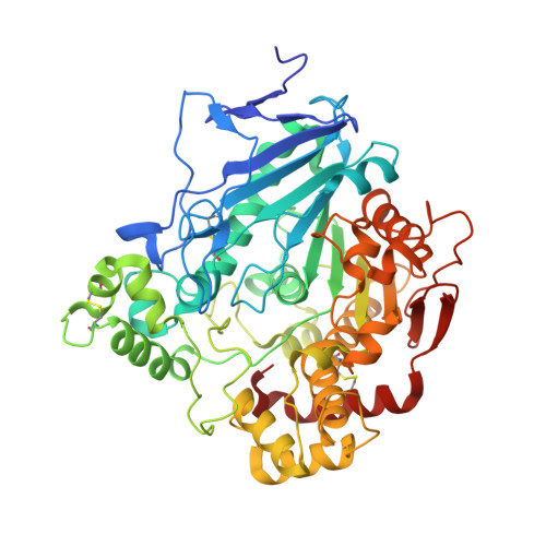
    From the fungus Aspergillus niger, we identified a new gene encoding protein EstA, a member of the alpha/beta-hydrolase fold superfamily but of unknown substrate specificity. EstA was overexpressed and its crystal structure was solved by molecular replacement using a lipase-acetylcholinesterase chimera template. The 2.1 A resolution structure of EstA reveals a canonical Ser/Glu/His catalytic triad located in a small pocket at the bottom of a large solvent-accessible, bowl-shaped cavity. Potential substrates selected by manual docking procedures were assayed for EstA activity. Consistent with the pocket geometry, preference for hydrolysis of short acyl/propyl chain substrates was found. Identification of close homologs from the genome of other fungi, of which some are broad host-range pathogens, defines EstA as the first member of a novel class of fungal esterases within the superfamily. Hence the structure of EstA constitutes a lead template in the design of new antifungal agents directed toward its pathogenic homologs.


  • Organizational Affiliation

    Architecture et Fonction des Macromolécules Biologiques, CNRS UMR-6098, 31 Chemin Joseph Aiguier, F-13402 Marseille Cedex 20, France. yves@afmb.cnrs-mrs.fr


Macromolecules
Find similar proteins by:  (by identity cutoff)  |  3D Structure
Entity ID: 1
MoleculeChains Sequence LengthOrganismDetailsImage
EstA
A, B
522Aspergillus nigerMutation(s): 0 
Gene Names: EstA
EC: 3.1.1
UniProt
Find proteins for Q6ED33 (Aspergillus niger)
Explore Q6ED33 
Go to UniProtKB:  Q6ED33
Entity Groups  
Sequence Clusters30% Identity50% Identity70% Identity90% Identity95% Identity100% Identity
UniProt GroupQ6ED33
Glycosylation
Glycosylation Sites: 4
Sequence Annotations
Expand
  • Reference Sequence
Oligosaccharides

Help

Entity ID: 2
MoleculeChains Length2D Diagram Glycosylation3D Interactions
alpha-D-mannopyranose-(1-4)-2-acetamido-2-deoxy-beta-D-glucopyranose-(1-4)-2-acetamido-2-deoxy-beta-D-glucopyranose
C
3N-Glycosylation
Glycosylation Resources
GlyTouCan:  G62182OO
GlyCosmos:  G62182OO
GlyGen:  G62182OO
Entity ID: 3
MoleculeChains Length2D Diagram Glycosylation3D Interactions
2-acetamido-2-deoxy-beta-D-glucopyranose-(1-4)-2-acetamido-2-deoxy-beta-D-glucopyranose
D
2N-Glycosylation
Glycosylation Resources
GlyTouCan:  G42666HT
GlyCosmos:  G42666HT
GlyGen:  G42666HT
Small Molecules
Ligands 5 Unique
IDChains Name / Formula / InChI Key2D Diagram3D Interactions
NAG
Query on NAG

Download Ideal Coordinates CCD File 
E [auth A]
F [auth A]
I [auth A]
J [auth A]
K [auth A]
2-acetamido-2-deoxy-beta-D-glucopyranose
C8 H15 N O6
OVRNDRQMDRJTHS-FMDGEEDCSA-N
MAN
Query on MAN

Download Ideal Coordinates CCD File 
G [auth A],
H [auth A],
S [auth B],
T [auth B],
U [auth B]
alpha-D-mannopyranose
C6 H12 O6
WQZGKKKJIJFFOK-PQMKYFCFSA-N
SO4
Query on SO4

Download Ideal Coordinates CCD File 
L [auth A],
Y [auth B]
SULFATE ION
O4 S
QAOWNCQODCNURD-UHFFFAOYSA-L
EDO
Query on EDO

Download Ideal Coordinates CCD File 
N [auth A],
O [auth A],
P [auth A],
Z [auth B]
1,2-ETHANEDIOL
C2 H6 O2
LYCAIKOWRPUZTN-UHFFFAOYSA-N
CL
Query on CL

Download Ideal Coordinates CCD File 
M [auth A]CHLORIDE ION
Cl
VEXZGXHMUGYJMC-UHFFFAOYSA-M
Experimental Data & Validation

Experimental Data

  • Method: X-RAY DIFFRACTION
  • Resolution: 2.10 Å
  • R-Value Free:  0.184 (Depositor), 0.190 (DCC) 
  • R-Value Work:  0.158 (Depositor), 0.170 (DCC) 
  • R-Value Observed: 0.158 (Depositor) 
Space Group: P 63
Unit Cell:
Length ( Å )Angle ( ˚ )
a = 167.489α = 90
b = 167.489β = 90
c = 112.827γ = 120
Software Package:
Software NamePurpose
REFMACrefinement
DENZOdata reduction
CCP4data scaling
AMoREphasing

Structure Validation

View Full Validation Report


Ligand Structure Quality Assessment 

Created with Raphaël 2.3.0Worse 01 BetterLigand structure goodness of fit to experimental dataBest fitted NAGClick on this verticalbar to view detailsBest fitted MANClick on this verticalbar to view details

Entry History 

Deposition Data

Revision History  (Full details and data files)

  • Version 1.0: 2004-07-27
    Type: Initial release
  • Version 1.1: 2008-04-27
    Changes: Version format compliance
  • Version 1.2: 2011-07-13
    Changes: Advisory, Version format compliance
  • Version 2.0: 2020-07-29
    Type: Remediation
    Reason: Carbohydrate remediation
    Changes: Advisory, Atomic model, Data collection, Derived calculations, Structure summary
  • Version 2.1: 2023-10-25
    Changes: Data collection, Database references, Refinement description, Structure summary
  • Version 2.2: 2024-10-23
    Changes: Structure summary