1V9Z | pdb_00001v9z

Crystal Structure of the heme PAS sensor domain of Ec DOS (Ferrous Form)


Experimental Data Snapshot

  • Method: X-RAY DIFFRACTION
  • Resolution: 1.90 Å
  • R-Value Free: 
    0.287 (Depositor), 0.280 (DCC) 
  • R-Value Work: 
    0.244 (Depositor), 0.240 (DCC) 

wwPDB Validation   3D Report Full Report


Ligand Structure Quality Assessment 

Created with Raphaël 2.3.0Worse 01 BetterLigand structure goodness of fit to experimental dataBest fitted HEMClick on this verticalbar to view details

This is version 1.3 of the entry. See complete history


Literature

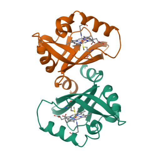
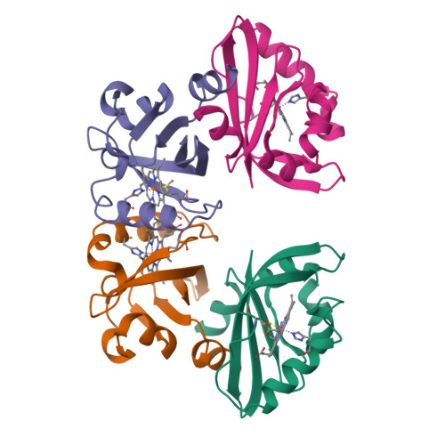
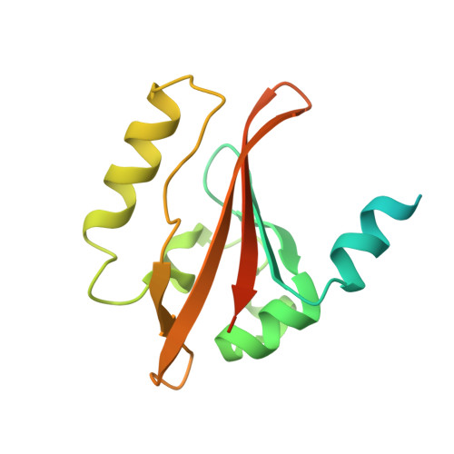
A redox-controlled molecular switch revealed by the crystal structure of a bacterial heme PAS sensor.

Kurokawa, H.Lee, D.S.Watanabe, M.Sagami, I.Mikami, B.Raman, C.S.Shimizu, T.

(2004) J Biological Chem 279: 20186-20193

  • DOI: https://doi.org/10.1074/jbc.M314199200
  • Primary Citation of Related Structures:  
    1V9Y, 1V9Z

  • PubMed Abstract: 

    PAS domains, which have been identified in over 1100 proteins from all three kingdoms of life, convert various input stimuli into signals that propagate to downstream components by modifying protein-protein interactions. One such protein is the Escherichia coli redox sensor, Ec DOS, a phosphodiesterase that degrades cyclic adenosine monophosphate in a redox-dependent manner. Here we report the crystal structures of the heme PAS domain of Ec DOS in both inactive Fe(3+) and active Fe(2+) forms at 1.32 and 1.9 A resolution, respectively. The protein folds into a characteristic PAS domain structure and forms a homodimer. In the Fe(3+) form, the heme iron is ligated to a His-77 side chain and a water molecule. Heme iron reduction is accompanied by heme-ligand switching from the water molecule to a side chain of Met-95 from the FG loop. Concomitantly, the flexible FG loop is significantly rigidified, along with a change in the hydrogen bonding pattern and rotation of subunits relative to each other. The present data led us to propose a novel redox-regulated molecular switch in which local heme-ligand switching may trigger a global "scissor-type" subunit movement that facilitates catalytic control.


  • Organizational Affiliation

    Institute of Multidisciplinary Research for Advanced Materials, Tohoku University, 2-1-1 Katahira, Aoba-ku, Sendai, Miyagi 980-8577, Japan. kurokawa@tagen.tohoku.ac.jp


Macromolecules
Find similar proteins by:  (by identity cutoff)  |  3D Structure
Entity ID: 1
MoleculeChains Sequence LengthOrganismDetailsImage
Heme pas sensor protein
A, B
167Escherichia coli K-12Mutation(s): 0 
Gene Names: DOS
EC: 3.1.4.52
UniProt
Find proteins for P76129 (Escherichia coli (strain K12))
Explore P76129 
Go to UniProtKB:  P76129
Entity Groups  
Sequence Clusters30% Identity50% Identity70% Identity90% Identity95% Identity100% Identity
UniProt GroupP76129
Sequence Annotations
Expand
  • Reference Sequence
Experimental Data & Validation

Experimental Data

  • Method: X-RAY DIFFRACTION
  • Resolution: 1.90 Å
  • R-Value Free:  0.287 (Depositor), 0.280 (DCC) 
  • R-Value Work:  0.244 (Depositor), 0.240 (DCC) 
Space Group: P 21 21 21
Unit Cell:
Length ( Å )Angle ( ˚ )
a = 46.4α = 90
b = 69.18β = 90
c = 82.45γ = 90
Software Package:
Software NamePurpose
CNSrefinement
MOSFLMdata reduction
CCP4data scaling
CNSphasing

Structure Validation

View Full Validation Report


Ligand Structure Quality Assessment 

Created with Raphaël 2.3.0Worse 01 BetterLigand structure goodness of fit to experimental dataBest fitted HEMClick on this verticalbar to view details

Entry History 

Deposition Data

Revision History  (Full details and data files)

  • Version 1.0: 2004-05-25
    Type: Initial release
  • Version 1.1: 2008-04-27
    Changes: Version format compliance
  • Version 1.2: 2011-07-13
    Changes: Source and taxonomy, Version format compliance
  • Version 1.3: 2023-12-27
    Changes: Data collection, Database references, Derived calculations