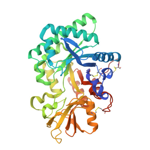
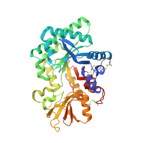
The crystal structure of Ym1 at 1.31 A resolution
Tsai, M.L., Liaw, S.H., Chang, N.C.(2004) J Struct Biol 148: 290-296
- PubMed: 15522777
- DOI: https://doi.org/10.1016/j.jsb.2004.07.002
- Primary Citation of Related Structures:
1VF8 - PubMed Abstract:
Upon nematode infection, murine peritoneal macrophages synthesize and secrete large amounts of the Ym1 protein, which is a unique functional marker for alternatively activated macrophages in T(H)2-mediated inflammatory responses. Ym1 shares significant structural similarity to the family 18 chitinases. Previously, Ym1 has been studied with respect to its carbohydrate-binding ability and glycosyl hydrolysis activity and this has led to various inconclusive interpretations. Our present co-crystallization and soaking experiments with various glucosamine or N-acetylglucosamine oligomers yield only the uncomplexed Ym1. The refined Ym1 structure at 1.31A resolution clearly displays a water cluster forming an extensive hydrogen bond network with the "active-site" residues. This water cluster contributes notable electron density to lower resolution maps and this might have misled and given rise to a previous proposal for a monoglucosamine-binding site for Ym1. A structural comparison of family 18 glycosidase (-like) proteins reveals a lack of several conserved residues in Ym1, and illustrates the versatility of the divergent active sites. Therefore, Ym1 may lack N-acetylglucosamine-binding affinity, and this suggests that a new direction should be taken to unravel the function of Ym1.
Organizational Affiliation:
Bioinformatics Program, National Yang-Ming University, Taipei 11221, Taiwan, ROC.