1WVG | pdb_00001wvg

Structure of CDP-D-glucose 4,6-dehydratase from Salmonella typhi


Experimental Data Snapshot

  • Method: X-RAY DIFFRACTION
  • Resolution: 1.80 Å
  • R-Value Free: 
    0.270 (Depositor) 
  • R-Value Work: 
    0.201 (Depositor), 0.200 (DCC) 
  • R-Value Observed: 
    0.203 (Depositor) 

Starting Model: experimental
View more details

wwPDB Validation   3D Report Full Report


Ligand Structure Quality Assessment 

Created with Raphaël 2.3.0Worse 01 BetterLigand structure goodness of fit to experimental dataBest fitted APRClick on this verticalbar to view detailsBest fitted CXYClick on this verticalbar to view details

This is version 1.4 of the entry. See complete history


Literature

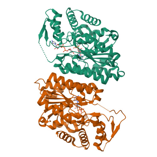
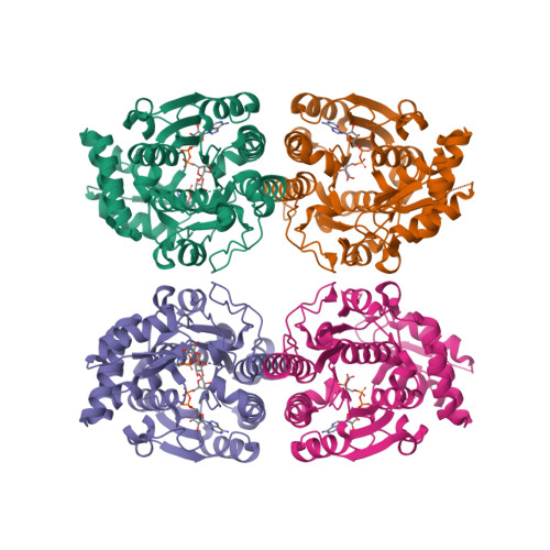
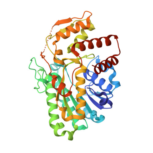
Structure of CDP-D-glucose 4,6-dehydratase from Salmonella typhi complexed with CDP-D-xylose.

Koropatkin, N.M.Holden, H.M.

(2005) Acta Crystallogr D Biol Crystallogr 61: 365-373

  • DOI: https://doi.org/10.1107/S0907444904033876
  • Primary Citation of Related Structures:  
    1WVG

  • PubMed Abstract: 

    Tyvelose is a unique 3,6-dideoxyhexose found in the O antigens of some pathogenic species of Yersinia and Salmonella. It is produced via a complex biochemical pathway that employs CDP-D-glucose as the starting ligand. CDP-D-glucose 4,6-dehydratase catalyzes the first irreversible step in the synthesis of this 3,6-dideoxysugar by converting CDP-D-glucose to CDP-4-keto-6-deoxyglucose via an NAD+ -dependent intramolecular oxidation-reduction reaction. Here, the cloning, protein purification and X-ray crystallographic analysis of CDP-D-glucose 4,6-dehydratase from Salmonella typhi complexed with the substrate analog CDP-D-xylose are described. Each subunit of the tetrameric enzyme folds into two domains. The N-terminal region contains a Rossmann fold and provides the platform for NAD(H) binding. The C-terminal motif is primarily composed of alpha-helices and houses the binding pocket for the CDP portion of the CDP-D-xylose ligand. The xylose moiety extends into the active-site cleft that is located between the two domains. Key residues involved in anchoring the sugar group to the protein include Ser134, Tyr159, Asn197 and Arg208. Strikingly, Ser134 O gamma and Tyr159 O eta sit within 2.9 A of the 4'-hydroxyl group of xylose. Additionally, the side chains of Asp135 and Lys136 are located at 3.5 and 3.2 A, respectively, from C-5 of xylose. In the structurally related dTDP-D-glucose 4,6-dehydratase, the Asp/Lys pair is replaced with an Asp/Glu couple. On the basis of this investigation, it can be speculated that Tyr159 serves as the catalytic base to abstract the 4'-hydroxyl proton from the sugar and that Asp135 and Lys136 play critical roles in the subsequent dehydration step that leads to the final product.


  • Organizational Affiliation

    Department of Biochemistry, University of Wisconsin, Madison, Wisconsin 53706-1544, USA.


Macromolecules
Find similar proteins by:  (by identity cutoff)  |  3D Structure
Entity ID: 1
MoleculeChains Sequence LengthOrganismDetailsImage
CDP-glucose 4,6-dehydratase
A, B
359Salmonella enterica subsp. enterica serovar Typhi str. CT18Mutation(s): 0 
Gene Names: ddhB
EC: 4.2.1.45
UniProt
Find proteins for P26397 (Salmonella typhimurium (strain LT2 / SGSC1412 / ATCC 700720))
Explore P26397 
Go to UniProtKB:  P26397
Entity Groups  
Sequence Clusters30% Identity50% Identity70% Identity90% Identity95% Identity100% Identity
UniProt GroupP26397
Sequence Annotations
Expand
  • Reference Sequence
Experimental Data & Validation

Experimental Data

  • Method: X-RAY DIFFRACTION
  • Resolution: 1.80 Å
  • R-Value Free:  0.270 (Depositor) 
  • R-Value Work:  0.201 (Depositor), 0.200 (DCC) 
  • R-Value Observed: 0.203 (Depositor) 
Space Group: P 31 2 1
Unit Cell:
Length ( Å )Angle ( ˚ )
a = 93.9α = 90
b = 93.9β = 90
c = 152.8γ = 120
Software Package:
Software NamePurpose
HKL-2000data collection
SCALEPACKdata scaling
AMoREphasing
TNTrefinement
HKL-2000data reduction

Structure Validation

View Full Validation Report


Ligand Structure Quality Assessment 

Created with Raphaël 2.3.0Worse 01 BetterLigand structure goodness of fit to experimental dataBest fitted APRClick on this verticalbar to view detailsBest fitted CXYClick on this verticalbar to view details

Entry History 

Deposition Data

Revision History  (Full details and data files)

  • Version 1.0: 2005-01-26
    Type: Initial release
  • Version 1.1: 2008-04-30
    Changes: Version format compliance
  • Version 1.2: 2011-07-13
    Changes: Source and taxonomy, Version format compliance
  • Version 1.3: 2024-03-13
    Changes: Data collection, Database references, Derived calculations
  • Version 1.4: 2024-04-03
    Changes: Refinement description