2BFY | pdb_00002bfy

Complex of Aurora-B with INCENP and Hesperadin.


Experimental Data Snapshot

  • Method: X-RAY DIFFRACTION
  • Resolution: 1.80 Å
  • R-Value Free: 
    0.226 (Depositor), 0.220 (DCC) 
  • R-Value Work: 
    0.203 (Depositor), 0.190 (DCC) 
  • R-Value Observed: 
    0.203 (Depositor) 

Starting Model: experimental
View more details

wwPDB Validation   3D Report Full Report


Ligand Structure Quality Assessment 

Created with Raphaël 2.3.0Worse 01 BetterLigand structure goodness of fit to experimental dataBest fitted H1NClick on this verticalbar to view details

This is version 1.4 of the entry. See complete history


Literature

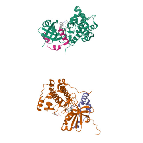
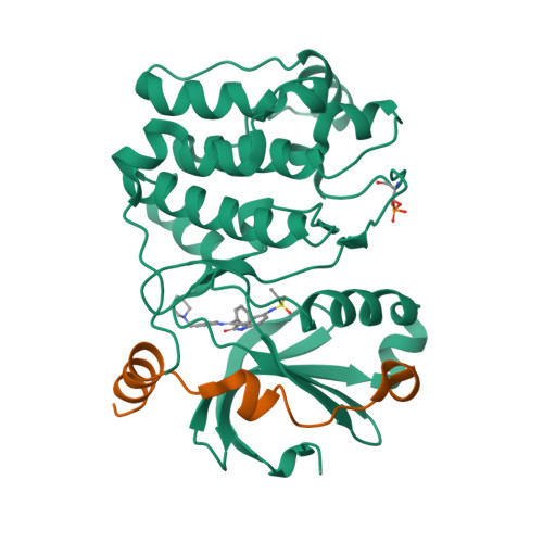
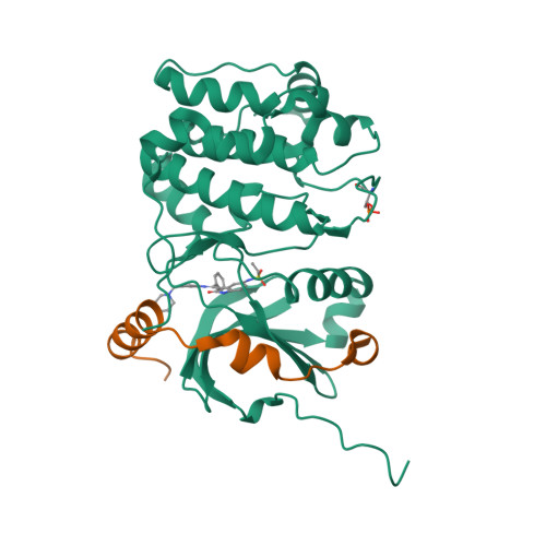
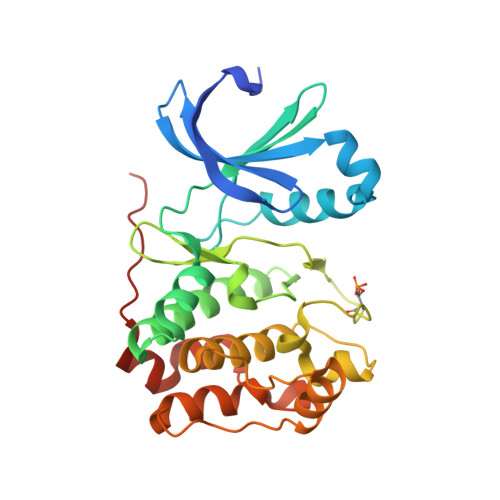
Mechanism of Aurora B Activation by Incenp and Inhibition by Hesperadin

Sessa, F.Mapelli, M.Ciferri, C.Tarricone, C.Areces, L.B.Schneider, T.R.Stukenberg, P.T.Musacchio, A.

(2005) Mol Cell 18: 379

  • DOI: https://doi.org/10.1016/j.molcel.2005.03.031
  • Primary Citation of Related Structures:  
    2BFX, 2BFY

  • PubMed Abstract: 

    Aurora family serine/threonine kinases control mitotic progression, and their deregulation is implicated in tumorigenesis. Aurora A and Aurora B, the best-characterized members of mammalian Aurora kinases, are approximately 60% identical but bind to unrelated activating subunits. The structure of the complex of Aurora A with the TPX2 activator has been reported previously. Here, we report the crystal structure of Aurora B in complex with the IN-box segment of the inner centromere protein (INCENP) activator and with the small molecule inhibitor Hesperadin. The Aurora B:INCENP complex is remarkably different from the Aurora A:TPX2 complex. INCENP forms a crown around the small lobe of Aurora B and induces the active conformation of the T loop allosterically. The structure represents an intermediate state of activation of Aurora B in which the Aurora B C-terminal segment stabilizes an open conformation of the catalytic cleft, and a critical ion pair in the kinase active site is impaired. Phosphorylation of two serines in the carboxyl terminus of INCENP generates the fully active kinase.


  • Organizational Affiliation

    Department of Experimental Oncology, European Institute of Oncology, Via Ripamonti 435, 20141 Milan, Italy.


Macromolecules
Find similar proteins by:  (by identity cutoff)  |  3D Structure
Entity ID: 1
MoleculeChains Sequence LengthOrganismDetailsImage
AURORA KINASE B-A
A, B
284Xenopus laevisMutation(s): 1 
EC: 2.7.11.1
UniProt
Find proteins for Q6DE08 (Xenopus laevis)
Explore Q6DE08 
Go to UniProtKB:  Q6DE08
Entity Groups  
Sequence Clusters30% Identity50% Identity70% Identity90% Identity95% Identity100% Identity
UniProt GroupQ6DE08
Sequence Annotations
Expand
  • Reference Sequence
Find similar proteins by:  (by identity cutoff)  |  3D Structure
Entity ID: 2
MoleculeChains Sequence LengthOrganismDetailsImage
INNER CENTROMERE PROTEIN A
C, D
43Xenopus laevisMutation(s): 0 
UniProt
Find proteins for O13024 (Xenopus laevis)
Explore O13024 
Go to UniProtKB:  O13024
Entity Groups  
Sequence Clusters30% Identity50% Identity70% Identity90% Identity95% Identity100% Identity
UniProt GroupO13024
Sequence Annotations
Expand
  • Reference Sequence
Small Molecules
Ligands 1 Unique
IDChains Name / Formula / InChI Key2D Diagram3D Interactions
H1N
Query on H1N

Download Ideal Coordinates CCD File 
E [auth A],
F [auth B]
N-[2-OXO-3-((E)-PHENYL{[4-(PIPERIDIN-1-YLMETHYL)PHENYL]IMINO}METHYL)-2,6-DIHYDRO-1H-INDOL-5-YL]ETHANESULFONAMIDE
C29 H32 N4 O3 S
SGZZQKMOFHIDKW-MCKMWFOCSA-N
Modified Residues  1 Unique
IDChains TypeFormula2D DiagramParent
TPO
Query on TPO
A, B
L-PEPTIDE LINKINGC4 H10 N O6 PTHR
Binding Affinity Annotations 
IDSourceBinding Affinity
H1N BindingDB:  2BFY IC50: 3 (nM) from 1 assay(s)
Experimental Data & Validation

Experimental Data

  • Method: X-RAY DIFFRACTION
  • Resolution: 1.80 Å
  • R-Value Free:  0.226 (Depositor), 0.220 (DCC) 
  • R-Value Work:  0.203 (Depositor), 0.190 (DCC) 
  • R-Value Observed: 0.203 (Depositor) 
Space Group: P 1 21 1
Unit Cell:
Length ( Å )Angle ( ˚ )
a = 45.945α = 90
b = 67.044β = 96.51
c = 116.462γ = 90
Software Package:
Software NamePurpose
CNSrefinement
HKL-2000data reduction
SCALEPACKdata scaling
AMoREphasing

Structure Validation

View Full Validation Report


Ligand Structure Quality Assessment 

Created with Raphaël 2.3.0Worse 01 BetterLigand structure goodness of fit to experimental dataBest fitted H1NClick on this verticalbar to view details

Entry History 

Deposition Data

Revision History  (Full details and data files)

  • Version 1.0: 2005-05-03
    Type: Initial release
  • Version 1.1: 2014-05-14
    Changes: Database references, Derived calculations, Non-polymer description, Other, Refinement description, Source and taxonomy, Structure summary, Version format compliance
  • Version 1.2: 2015-05-20
    Changes: Structure summary
  • Version 1.3: 2023-12-13
    Changes: Data collection, Database references, Derived calculations, Other, Refinement description
  • Version 1.4: 2024-11-06
    Changes: Structure summary