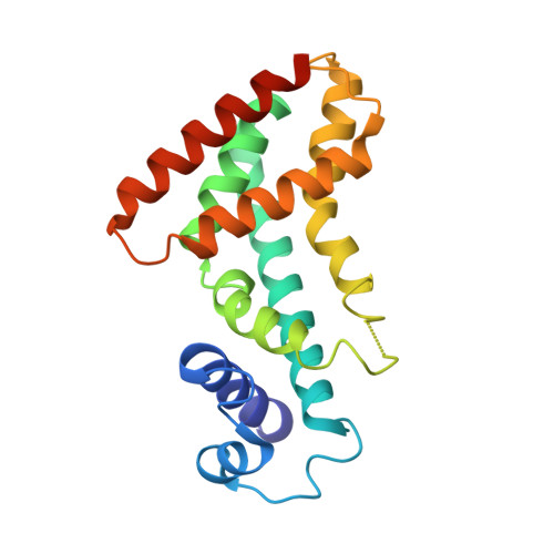
2HQ5

Crystal structure of multidrug binding protein QacR from Staphylococcus aureus cocrystallized with compound DB359


Experimental Data Snapshot

  • Method: X-RAY DIFFRACTION
  • Resolution: 2.80 Å
  • R-Value Free: 0.259 
  • R-Value Work: 0.213 
  • R-Value Observed: 0.213 

Starting Model: experimental
View more details

wwPDB Validation   3D Report Full Report


This is version 1.4 of the entry. See complete history


Literature

Multidrug-Binding Transcription Factor QacR Binds the Bivalent Aromatic Diamidines DB75 and DB359 in Multiple Positions

Brooks, B.E.Piro, K.M.Brennan, R.G.

(2007) J Am Chem Soc 129: 8389-8395

  • DOI: https://doi.org/10.1021/ja072576v
  • Primary Citation of Related Structures:  
    2DTZ, 2HQ5

  • PubMed Abstract: 

    Staphylococcus aureus QacR is a multidrug-binding transcription repressor. Crystal structures of multiple QacR-drug complexes reveal that these toxins bind in a large pocket, which is composed of smaller overlapping "minipockets". Stacking, van der Waals, and ionic interactions are common features of binding, whereas hydrogen bonds are limited. Pentamidine, a bivalent aromatic diamidine, interacts with QacR differently as one positively charged benzamidine moiety is neutralized by the dipoles of side-chain and peptide backbone oxygens rather than a formal negative charge from proximal acidic residues. To understand the binding mechanisms of other bivalent benzamidines, we determined the crystal structures of the QacR-DB75 and QacR-DB359 complexes and measured their binding affinities. Although these rigid aromatic diamidines bind with low-micromolar affinities, they do not use single, discrete binding modes. Such promiscuous binding underscores the intrinsic chemical redundancy of the QacR multidrug-binding pocket. Chemical redundancy is likely a hallmark of all multidrug-binding pockets, yet it is utilized by only a subset of drugs, which, for QacR, so far appears to be limited to chemically rigid, bivalent compounds.


  • Organizational Affiliation

    Department of Biochemistry and Molecular Biology, University of Texas MD Anderson Cancer Center, Unit 1000, Houston, TX 77030, USA.


Macromolecules
Find similar proteins by:  (by identity cutoff)  |  3D Structure
Entity ID: 1
MoleculeChains Sequence LengthOrganismDetailsImage
HTH-type transcriptional regulator qacRA [auth B],
B [auth D],
C [auth A],
D [auth E]
194Staphylococcus aureusMutation(s): 2 
Gene Names: qacRSAVP031
UniProt
Find proteins for P0A0N4 (Staphylococcus aureus)
Explore P0A0N4 
Go to UniProtKB:  P0A0N4
Entity Groups  
Sequence Clusters30% Identity50% Identity70% Identity90% Identity95% Identity100% Identity
UniProt GroupP0A0N4
Sequence Annotations
Expand
  • Reference Sequence
Small Molecules
Ligands 1 Unique
IDChains Name / Formula / InChI Key2D Diagram3D Interactions
SO4
Query on SO4

Download Ideal Coordinates CCD File 
E [auth B]
F [auth D]
G [auth D]
H [auth D]
I [auth A]
E [auth B],
F [auth D],
G [auth D],
H [auth D],
I [auth A],
J [auth A],
K [auth A],
L [auth A],
M [auth A],
N [auth A],
O [auth E],
P [auth E],
Q [auth E],
R [auth E]
SULFATE ION
O4 S
QAOWNCQODCNURD-UHFFFAOYSA-L
Experimental Data & Validation

Experimental Data

  • Method: X-RAY DIFFRACTION
  • Resolution: 2.80 Å
  • R-Value Free: 0.259 
  • R-Value Work: 0.213 
  • R-Value Observed: 0.213 
  • Space Group: P 42 21 2
Unit Cell:
Length ( Å )Angle ( ˚ )
a = 171.366α = 90
b = 171.366β = 90
c = 94.496γ = 90
Software Package:
Software NamePurpose
CNSrefinement
MOSFLMdata reduction
SCALAdata scaling
MOLREPphasing

Structure Validation

View Full Validation Report


Entry History 

Deposition Data

Revision History  (Full details and data files)

  • Version 1.0: 2007-07-17
    Type: Initial release
  • Version 1.1: 2008-05-01
    Changes: Version format compliance
  • Version 1.2: 2011-07-13
    Changes: Version format compliance
  • Version 1.3: 2021-11-10
    Changes: Database references, Derived calculations
  • Version 1.4: 2023-10-25
    Changes: Data collection, Refinement description