2JJA | pdb_00002jja

Crystal structure of GNA with synthetic copper base pair


Experimental Data Snapshot

  • Method: X-RAY DIFFRACTION
  • Resolution: 1.30 Å
  • R-Value Free: 
    0.148 (Depositor), 0.150 (DCC) 
  • R-Value Work: 
    0.124 (Depositor), 0.130 (DCC) 
  • R-Value Observed: 
    0.126 (Depositor) 

wwPDB Validation   3D Report Full Report


This is version 2.1 of the entry. See complete history


Literature

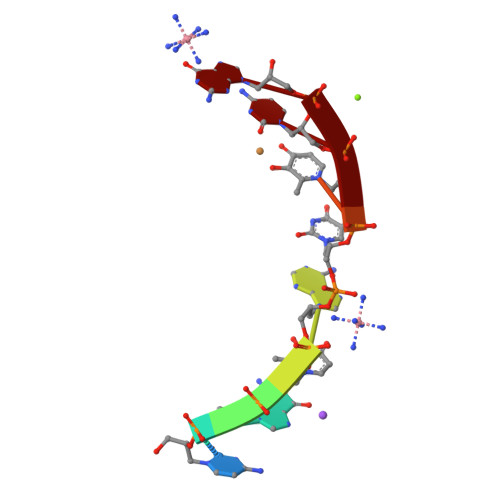
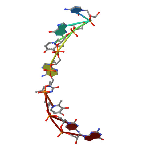
Duplex Structure of a Minimal Nucleic Acid.

Schlegel, M.K.Essen, L.-O.Meggers, E.

(2008) J Am Chem Soc 130: 8158

  • DOI: https://doi.org/10.1021/ja802788g
  • Primary Citation of Related Structures:  
    2JJA

  • PubMed Abstract: 

    The crystal structure of an 8-mer (S)-GNA duplex is presented. As a tool for phasing, the anomalous diffraction of two copper(II) ions within two artificial metallo-base pairs was employed. The duplex structure confirms a canonical Watson-Crick base pairing scheme of GNA with antiparallel strands. The duplex secondary structure is distinct from canonical A- and B-form nucleic acids and can be described as a right-handed helical ribbon wrapped around the helix axis, resulting in a large hollow core. Most intriguingly, neighboring base pairs slide strongly against each other, resulting in extensive interstrand base-base hydrophobic interactions along with unusual hydrophobic intrastrand interactions of nucleobases with their backbone. These results reveal how a minimal nucleic acid backbone can support highly stable Watson-Crick-like duplex formation.


  • Organizational Affiliation

    Fachbereich Chemie, Philipps-Universität Marburg, Hans-Meerwein Strasse, D-35043 Marburg, Germany.


Macromolecules

Find similar nucleic acids by:  Sequence   |   3D Structure  

Entity ID: 1
MoleculeChains LengthOrganismImage
GNA8synthetic construct
Sequence Annotations
Expand
  • Reference Sequence
Small Molecules
Ligands 4 Unique
IDChains Name / Formula / InChI Key2D Diagram3D Interactions
NCO
Query on NCO

Download Ideal Coordinates CCD File 
E [auth A],
F [auth A]
COBALT HEXAMMINE(III)
Co H18 N6
DYLMFCCYOUSRTK-UHFFFAOYSA-N
CU
Query on CU

Download Ideal Coordinates CCD File 
B [auth A]COPPER (II) ION
Cu
JPVYNHNXODAKFH-UHFFFAOYSA-N
MG
Query on MG

Download Ideal Coordinates CCD File 
C [auth A],
D [auth A]
MAGNESIUM ION
Mg
JLVVSXFLKOJNIY-UHFFFAOYSA-N
NA
Query on NA

Download Ideal Coordinates CCD File 
G [auth A]SODIUM ION
Na
FKNQFGJONOIPTF-UHFFFAOYSA-N
Experimental Data & Validation

Experimental Data

  • Method: X-RAY DIFFRACTION
  • Resolution: 1.30 Å
  • R-Value Free:  0.148 (Depositor), 0.150 (DCC) 
  • R-Value Work:  0.124 (Depositor), 0.130 (DCC) 
  • R-Value Observed: 0.126 (Depositor) 
Space Group: I 21 21 21
Unit Cell:
Length ( Å )Angle ( ˚ )
a = 18.16α = 90
b = 48.59β = 90
c = 59.87γ = 90
Software Package:
Software NamePurpose
REFMACrefinement
XDSdata reduction
XSCALEdata scaling

Structure Validation

View Full Validation Report


Entry History 

Deposition Data

Revision History  (Full details and data files)

  • Version 1.0: 2008-07-22
    Type: Initial release
  • Version 1.1: 2011-05-07
    Changes: Version format compliance
  • Version 1.2: 2011-07-13
    Changes: Version format compliance
  • Version 2.0: 2022-06-22
    Changes: Data collection, Database references, Derived calculations, Other, Polymer sequence
  • Version 2.1: 2024-06-19
    Changes: Data collection