PCS-based structure determination of protein-protein complexes
Saio, T., Yokochi, M., Kumeta, H., Inagaki, F.(2010) J Biomol NMR 46: 271-280
- PubMed: 20300805
- DOI: https://doi.org/10.1007/s10858-010-9401-4
- Primary Citation of Related Structures:
2KTR - PubMed Abstract:
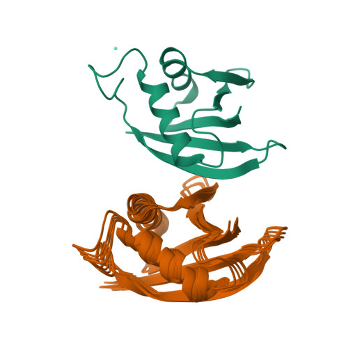
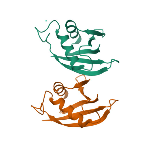
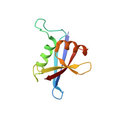
A simple and fast nuclear magnetic resonance method for docking proteins using pseudo-contact shift (PCS) and (1)H(N)/(15)N chemical shift perturbation is presented. PCS is induced by a paramagnetic lanthanide ion that is attached to a target protein using a lanthanide binding peptide tag anchored at two points. PCS provides long-range (approximately 40 A) distance and angular restraints between the lanthanide ion and the observed nuclei, while the (1)H(N)/(15)N chemical shift perturbation data provide loose contact-surface information. The usefulness of this method was demonstrated through the structure determination of the p62 PB1-PB1 complex, which forms a front-to-back 20 kDa homo-oligomer. As p62 PB1 does not intrinsically bind metal ions, the lanthanide binding peptide tag was attached to one subunit of the dimer at two anchoring points. Each monomer was treated as a rigid body and was docked based on the backbone PCS and backbone chemical shift perturbation data. Unlike NOE-based structural determination, this method only requires resonance assignments of the backbone (1)H(N)/(15)N signals and the PCS data obtained from several sets of two-dimensional (15)N-heteronuclear single quantum coherence spectra, thus facilitating rapid structure determination of the protein-protein complex.
Organizational Affiliation:
Graduate School of Life Science, Hokkaido University, Sapporo, 001-0021, Japan.