2O6P | pdb_00002o6p

Crystal Structure of the heme-IsdC complex


Experimental Data Snapshot

  • Method: X-RAY DIFFRACTION
  • Resolution: 1.50 Å
  • R-Value Free: 
    0.222 (Depositor), 0.220 (DCC) 
  • R-Value Work: 
    0.193 (Depositor), 0.190 (DCC) 
  • R-Value Observed: 
    0.195 (Depositor) 

wwPDB Validation   3D Report Full Report


Ligand Structure Quality Assessment 

Created with Raphaël 2.3.0Worse 01 BetterLigand structure goodness of fit to experimental dataBest fitted HEMClick on this verticalbar to view details

This is version 1.4 of the entry. See complete history


Literature

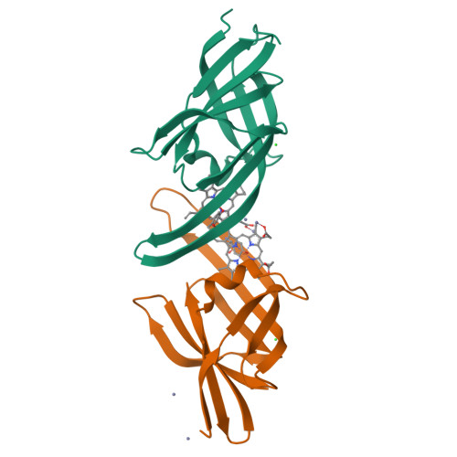
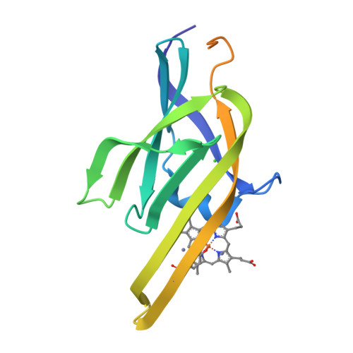
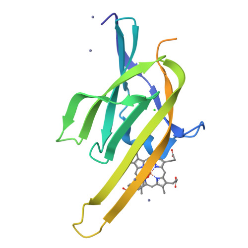
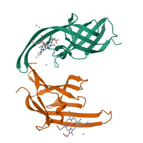
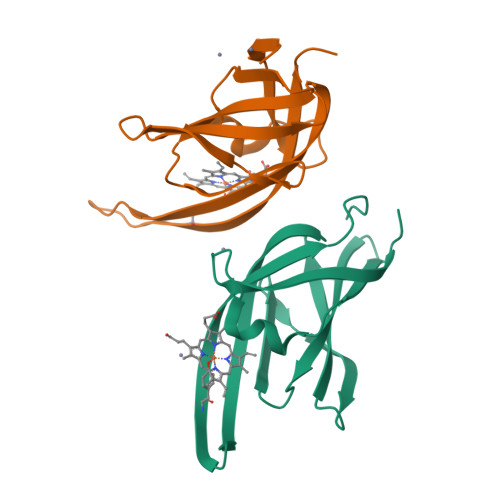
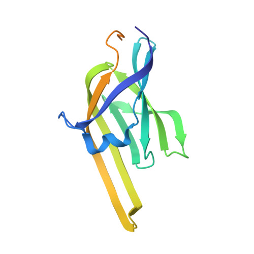
Crystal structure of the heme-IsdC complex, the central conduit of the Isd iron/heme uptake system in Staphylococcus aureus.

Sharp, K.H.Schneider, S.Cockayne, A.Paoli, M.

(2007) J Biological Chem 282: 10625-10631

  • DOI: https://doi.org/10.1074/jbc.M700234200
  • Primary Citation of Related Structures:  
    2O6P

  • PubMed Abstract: 

    Pathogens such as Staphylococcus aureus require iron to survive and have evolved specialized proteins to steal heme from their host. IsdC is the central conduit of the Isd (iron-regulated surface determinant) multicomponent heme uptake machinery; staphylococcal cell-surface proteins such as IsdA, IsdB, and IsdH are thought to funnel their molecular cargo to IsdC, which then mediates the transfer of the iron-containing nutrient to the membrane translocation system IsdDEF. The structure of the heme-IsdC complex reveals a novel heme site within an immunoglobulin-like domain and sheds light on its binding mechanism. The folding topology is reminiscent of the architecture of cytochrome f, cellobiose dehydrogenase, and ethylbenzene dehydrogenase; in these three proteins, the heme is bound in an equivalent position, but interestingly, IsdC features a distinct binding pocket with the ligand located next to the hydrophobic core of the beta-sandwich. The iron is coordinated with a tyrosine surrounded by several non-polar side chains that cluster into a tightly packed proximal side. On the other hand, the distal side is relatively exposed with a short helical peptide segment that acts as a lip clasping onto almost half of the porphyrin plane. This structural feature is argued to play a role in the mechanism of binding and release by switching to an open conformation and thus loosening the interactions holding the heme. The structure of the heme-IsdC complex provides a template for the understanding of other proteins, such as IsdA, IsdB, and IsdH, that contain the same heme-binding module as IsdC, known as the NEAT (near transporter) domain.


  • Organizational Affiliation

    School of Pharmacy, Centre for Biomolecular Sciences, University of Nottingham, University Park, Nottingham NG7 2RD, United Kingdom.


Macromolecules
Find similar proteins by:  (by identity cutoff)  |  3D Structure
Entity ID: 1
MoleculeChains Sequence LengthOrganismDetailsImage
Iron-regulated surface determinant protein C
A, B
161Staphylococcus aureus subsp. aureus N315Mutation(s): 0 
UniProt
Find proteins for Q8KQR1 (Staphylococcus aureus (strain NCTC 8325 / PS 47))
Explore Q8KQR1 
Go to UniProtKB:  Q8KQR1
Entity Groups  
Sequence Clusters30% Identity50% Identity70% Identity90% Identity95% Identity100% Identity
UniProt GroupQ8KQR1
Sequence Annotations
Expand
  • Reference Sequence
Small Molecules
Ligands 3 Unique
IDChains Name / Formula / InChI Key2D Diagram3D Interactions
HEM
Query on HEM

Download Ideal Coordinates CCD File 
F [auth A],
L [auth B]
PROTOPORPHYRIN IX CONTAINING FE
C34 H32 Fe N4 O4
KABFMIBPWCXCRK-RGGAHWMASA-L
ZN
Query on ZN

Download Ideal Coordinates CCD File 
C [auth A]
D [auth A]
G [auth B]
H [auth B]
I [auth B]
ZINC ION
Zn
PTFCDOFLOPIGGS-UHFFFAOYSA-N
CL
Query on CL

Download Ideal Coordinates CCD File 
E [auth A],
K [auth B]
CHLORIDE ION
Cl
VEXZGXHMUGYJMC-UHFFFAOYSA-M
Experimental Data & Validation

Experimental Data

  • Method: X-RAY DIFFRACTION
  • Resolution: 1.50 Å
  • R-Value Free:  0.222 (Depositor), 0.220 (DCC) 
  • R-Value Work:  0.193 (Depositor), 0.190 (DCC) 
  • R-Value Observed: 0.195 (Depositor) 
Space Group: P 21 21 21
Unit Cell:
Length ( Å )Angle ( ˚ )
a = 47.489α = 90
b = 70.441β = 90
c = 88.685γ = 90
Software Package:
Software NamePurpose
REFMACrefinement
MOSFLMdata reduction
CCP4data scaling
SHARPphasing

Structure Validation

View Full Validation Report


Ligand Structure Quality Assessment 

Created with Raphaël 2.3.0Worse 01 BetterLigand structure goodness of fit to experimental dataBest fitted HEMClick on this verticalbar to view details

Entry History 

Deposition Data

Revision History  (Full details and data files)

  • Version 1.0: 2007-02-06
    Type: Initial release
  • Version 1.1: 2008-05-01
    Changes: Version format compliance
  • Version 1.2: 2011-07-13
    Changes: Derived calculations, Source and taxonomy, Version format compliance
  • Version 1.3: 2017-12-13
    Changes: Advisory, Database references
  • Version 1.4: 2023-12-27
    Changes: Advisory, Data collection, Database references, Derived calculations