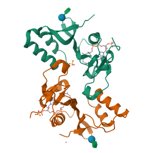
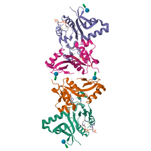
Crystal structure of the Bacillus subtilis N-acetyltransferase YlbP protein in complex with Coenzyme-A.
Minasov, G., Shuvalova, L., Kiryukhina, O., Vorontsov, I.I., Collart, F.R., Joachimiak, A., Anderson, W.F.To be published.
Experimental Data Snapshot
Entity ID: 1 | |||||
|---|---|---|---|---|---|
| Molecule | Chains | Sequence Length | Organism | Details | Image |
| Uncharacterized N-acetyltransferase ylbP | 163 | Bacillus subtilis | Mutation(s): 0 Gene Names: ylbP, BSU15100 EC: 2.3.1 | 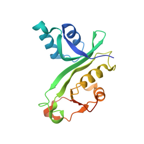 | |
UniProt | |||||
Find proteins for O34468 (Bacillus subtilis (strain 168)) Explore O34468 Go to UniProtKB: O34468 | |||||
Entity Groups | |||||
| Sequence Clusters | 30% Identity50% Identity70% Identity90% Identity95% Identity100% Identity | ||||
| UniProt Group | O34468 | ||||
Sequence AnnotationsExpand | |||||
| |||||
| Ligands 3 Unique | |||||
|---|---|---|---|---|---|
| ID | Chains | Name / Formula / InChI Key | 2D Diagram | 3D Interactions | |
| COA Query on COA | F [auth A], J [auth B] | COENZYME A C21 H36 N7 O16 P3 S RGJOEKWQDUBAIZ-IBOSZNHHSA-N |  | ||
| SO4 Query on SO4 | E [auth A], I [auth B] | SULFATE ION O4 S QAOWNCQODCNURD-UHFFFAOYSA-L |  | ||
| CO Query on CO | G [auth B], H [auth B] | COBALT (II) ION Co XLJKHNWPARRRJB-UHFFFAOYSA-N |  | ||
Entity ID: 2 | |||||
|---|---|---|---|---|---|
| ID | Chains | Name | Type/Class | 2D Diagram | 3D Interactions |
| PRD_900003 Query on PRD_900003 | C, D | sucrose | Oligosaccharide / Nutrient |  | |
| Length ( Å ) | Angle ( ˚ ) |
|---|---|
| a = 148.572 | α = 90 |
| b = 148.572 | β = 90 |
| c = 102.29 | γ = 120 |
| Software Name | Purpose |
|---|---|
| REFMAC | refinement |
| Blu-Ice | data collection |
| HKL-2000 | data reduction |
| HKL-2000 | data scaling |
| SHARP | phasing |
RCSB PDB is hosted by

RCSB PDB is a member of the