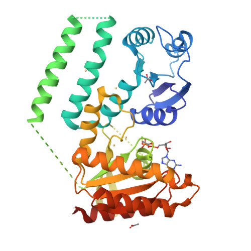
Crystal structure of a GTP-binding protein from the hyperthermophilic archaeon Sulfolobus solfataricus.
Wu, H., Sun, L., Brouns, S.J., Fu, S., Akerboom, J., Li, X., Zhang, C., Rao, Z., Van der Oost, J.To be published.
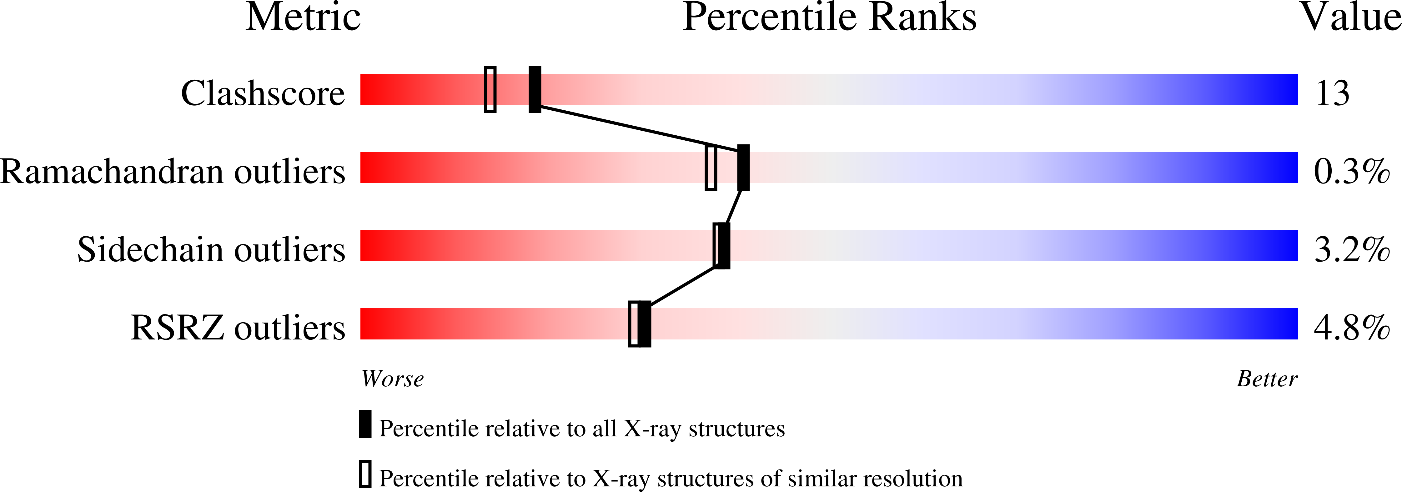
Experimental Data Snapshot
Entity ID: 1 | |||||
|---|---|---|---|---|---|
| Molecule | Chains | Sequence Length | Organism | Details | Image |
| GTP-binding protein | 364 | N/A | Mutation(s): 0 Gene Names: hflX, SSO0269 | 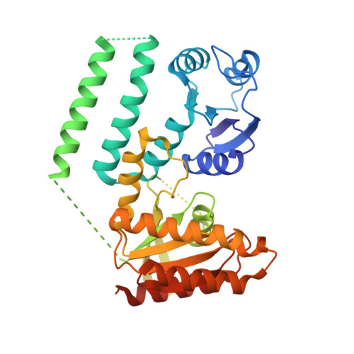 | |
UniProt | |||||
Find proteins for Q980M3 (Saccharolobus solfataricus (strain ATCC 35092 / DSM 1617 / JCM 11322 / P2)) Explore Q980M3 Go to UniProtKB: Q980M3 | |||||
Entity Groups | |||||
| Sequence Clusters | 30% Identity50% Identity70% Identity90% Identity95% Identity100% Identity | ||||
| UniProt Group | Q980M3 | ||||
Sequence AnnotationsExpand | |||||
| |||||
| Ligands 4 Unique | |||||
|---|---|---|---|---|---|
| ID | Chains | Name / Formula / InChI Key | 2D Diagram | 3D Interactions | |
| GDP Query on GDP | H [auth A] | GUANOSINE-5'-DIPHOSPHATE C10 H15 N5 O11 P2 QGWNDRXFNXRZMB-UUOKFMHZSA-N |  | ||
| CD Query on CD | B [auth A], C [auth A], D [auth A], E [auth A] | CADMIUM ION Cd WLZRMCYVCSSEQC-UHFFFAOYSA-N |  | ||
| ACT Query on ACT | G [auth A], I [auth A], J [auth A] | ACETATE ION C2 H3 O2 QTBSBXVTEAMEQO-UHFFFAOYSA-M |  | ||
| MG Query on MG | F [auth A] | MAGNESIUM ION Mg JLVVSXFLKOJNIY-UHFFFAOYSA-N |  | ||
| Length ( Å ) | Angle ( ˚ ) |
|---|---|
| a = 64.991 | α = 90 |
| b = 72.403 | β = 90 |
| c = 96.016 | γ = 90 |
| Software Name | Purpose |
|---|---|
| DENZO | data reduction |
| SCALEPACK | data scaling |
| REFMAC | refinement |
| PDB_EXTRACT | data extraction |
| CrystalClear | data collection |
| SHARP | phasing |
| DM | phasing |