2V52 | pdb_00002v52

Structure of MAL-RPEL2 complexed to G-actin


Experimental Data Snapshot

  • Method: X-RAY DIFFRACTION
  • Resolution: 1.45 Å
  • R-Value Free: 
    0.188 (Depositor), 0.180 (DCC) 
  • R-Value Work: 
    0.147 (Depositor), 0.140 (DCC) 
  • R-Value Observed: 
    0.149 (Depositor) 

wwPDB Validation   3D Report Full Report


Ligand Structure Quality Assessment 

Created with Raphaël 2.3.0Worse 01 BetterLigand structure goodness of fit to experimental dataBest fitted ATPClick on this verticalbar to view detailsBest fitted LABClick on this verticalbar to view details

This is version 2.1 of the entry. See complete history


Literature

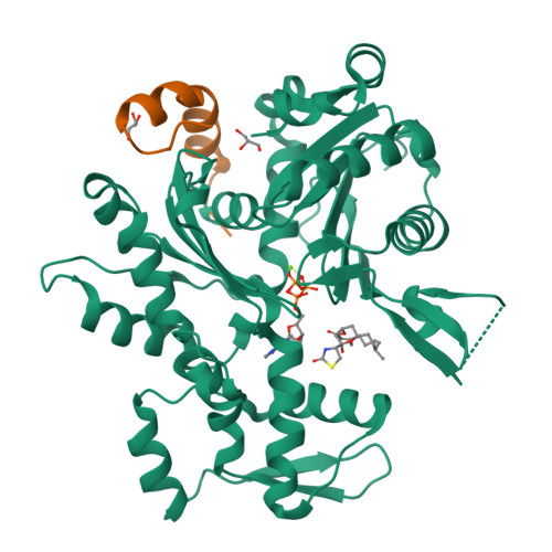
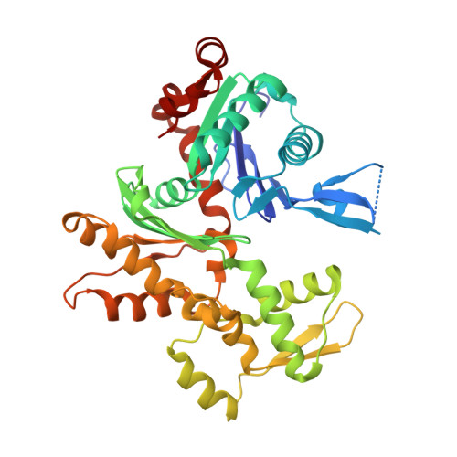
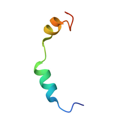
Molecular basis for G-actin binding to RPEL motifs from the serum response factor coactivator MAL.

Mouilleron, S.Guettler, S.Langer, C.A.Treisman, R.McDonald, N.Q.

(2008) EMBO J 27: 3198-3208

  • DOI: https://doi.org/10.1038/emboj.2008.235
  • Primary Citation of Related Structures:  
    2V51, 2V52

  • PubMed Abstract: 

    Serum response factor transcriptional activity is controlled through interactions with regulatory cofactors such as the coactivator MAL/MRTF-A (myocardin-related transcription factor A). MAL is itself regulated in vivo by changes in cellular actin dynamics, which alter its interaction with G-actin. The G-actin-sensing mechanism of MAL/MRTF-A resides in its N-terminal domain, which consists of three tandem RPEL repeats. We describe the first molecular insights into RPEL function obtained from structures of two independent RPEL(MAL) peptide:G-actin complexes. Both RPEL peptides bind to the G-actin hydrophobic cleft and to subdomain 3. These RPEL(MAL):G-actin structures explain the sequence conservation defining the RPEL motif, including the invariant arginine. Characterisation of the RPEL(MAL):G-actin interaction by fluorescence anisotropy and cell reporter-based assays validates the significance of actin-binding residues for proper MAL localisation and regulation in vivo. We identify important differences in G-actin engagement between the two RPEL(MAL) structures. Comparison with other actin-binding proteins reveals an unexpected similarity to the vitamin-D-binding protein, extending the G-actin-binding protein repertoire.


  • Organizational Affiliation

    Structural Biology Laboratory, Cancer Research UK, London Research Institute, London, UK.


Macromolecules
Find similar proteins by:  (by identity cutoff)  |  3D Structure
Entity ID: 1
MoleculeChains Sequence LengthOrganismDetailsImage
ACTIN, ALPHA SKELETAL MUSCLEA [auth B]377Oryctolagus cuniculusMutation(s): 0 
EC: 3.6.4
UniProt
Find proteins for P68135 (Oryctolagus cuniculus)
Explore P68135 
Go to UniProtKB:  P68135
Entity Groups  
Sequence Clusters30% Identity50% Identity70% Identity90% Identity95% Identity100% Identity
UniProt GroupP68135
Sequence Annotations
Expand
  • Reference Sequence
Find similar proteins by:  (by identity cutoff)  |  3D Structure
Entity ID: 2
MoleculeChains Sequence LengthOrganismDetailsImage
MKL/MYOCARDIN-LIKE PROTEIN 1B [auth M]32Mus musculusMutation(s): 0 
UniProt
Find proteins for Q8K4J6 (Mus musculus)
Explore Q8K4J6 
Go to UniProtKB:  Q8K4J6
Entity Groups  
Sequence Clusters30% Identity50% Identity70% Identity90% Identity95% Identity100% Identity
UniProt GroupQ8K4J6
Sequence Annotations
Expand
  • Reference Sequence
Small Molecules
Binding Affinity Annotations 
IDSourceBinding Affinity
LAB BindingDB:  2V52 IC50: 8470 (nM) from 1 assay(s)
Experimental Data & Validation

Experimental Data

  • Method: X-RAY DIFFRACTION
  • Resolution: 1.45 Å
  • R-Value Free:  0.188 (Depositor), 0.180 (DCC) 
  • R-Value Work:  0.147 (Depositor), 0.140 (DCC) 
  • R-Value Observed: 0.149 (Depositor) 
Space Group: P 21 21 21
Unit Cell:
Length ( Å )Angle ( ˚ )
a = 54.75α = 90
b = 55.44β = 90
c = 138.39γ = 90
Software Package:
Software NamePurpose
PHENIXrefinement
MOSFLMdata reduction
SCALAdata scaling
PHASERphasing

Structure Validation

View Full Validation Report


Ligand Structure Quality Assessment 

Created with Raphaël 2.3.0Worse 01 BetterLigand structure goodness of fit to experimental dataBest fitted ATPClick on this verticalbar to view detailsBest fitted LABClick on this verticalbar to view details

Entry History 

Deposition Data

Revision History  (Full details and data files)

  • Version 1.0: 2008-11-25
    Type: Initial release
  • Version 1.1: 2011-05-08
    Changes: Version format compliance
  • Version 1.2: 2011-07-13
    Changes: Version format compliance
  • Version 1.3: 2017-07-12
    Changes: Advisory, Derived calculations
  • Version 2.0: 2018-02-28
    Changes: Atomic model, Database references
  • Version 2.1: 2024-05-08
    Changes: Data collection, Database references, Derived calculations, Other