2WQ7 | pdb_00002wq7

Structure of the 6-4 photolyase of D. melanogaster in complex with the non-natural N4-methyl T(6-4)C lesion


Experimental Data Snapshot

  • Method: X-RAY DIFFRACTION
  • Resolution: 2.00 Å
  • R-Value Free: 
    0.209 (Depositor), 0.200 (DCC) 
  • R-Value Work: 
    0.178 (Depositor), 0.170 (DCC) 
  • R-Value Observed: 
    0.179 (Depositor) 

Starting Model: experimental
View more details

wwPDB Validation   3D Report Full Report


Ligand Structure Quality Assessment 

Created with Raphaël 2.3.0Worse 01 BetterLigand structure goodness of fit to experimental dataBest fitted FADClick on this verticalbar to view details

This is version 2.0 of the entry. See complete history


Literature

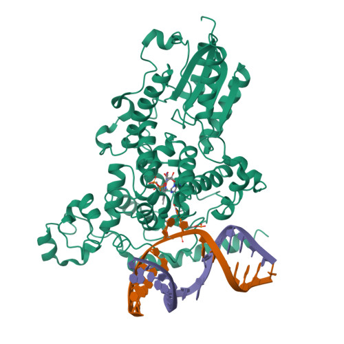
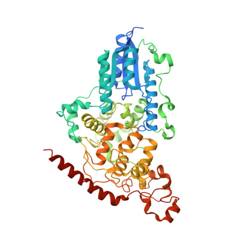
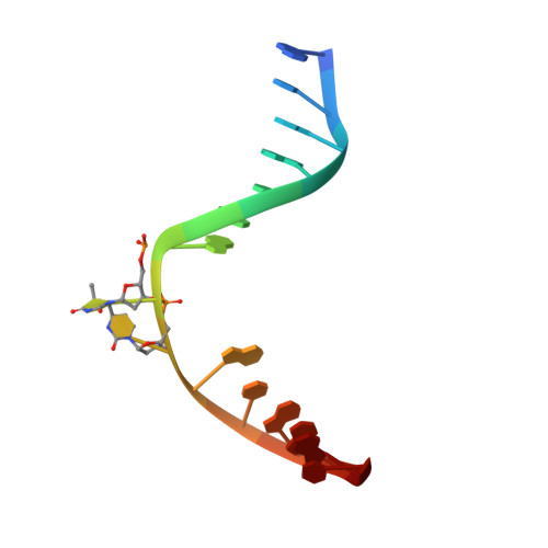
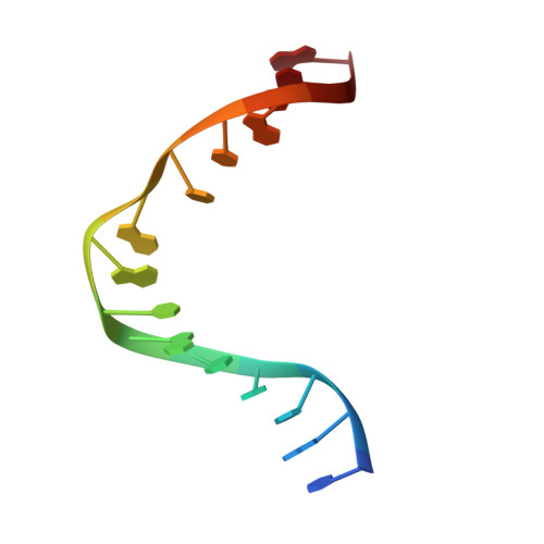
DNA (6-4) Photolyases Reduce Dewar Isomers for Isomerization Into (6-4) Lesions.

Glas, A.F.Kaya, E.Schneider, S.Heil, K.Fazio, D.Maul, M.J.Carell, T.

(2010) J Am Chem Soc 132: 3254

  • DOI: https://doi.org/10.1021/ja910917f
  • Primary Citation of Related Structures:  
    2WQ6, 2WQ7

  • PubMed Abstract: 

    Repair of the Dewar valence isomers by (6-4) photolyases proceeds via an enzyme catalyzed ring-opening reaction of the Dewar lesion to the (6-4) photoproduct.


  • Organizational Affiliation

    Center for Integrative Protein Science, Department of Chemistry and Biochemistry, Ludwig-Maximilians University Munich, Butenandtstr. 5-13, 81377 Munich, Germany.


Macromolecules

Find similar proteins by:  (by identity cutoff)  |  3D Structure
Entity ID: 1
MoleculeChains Sequence LengthOrganismDetailsImage
RE11660P543Drosophila melanogasterMutation(s): 0 
EC: 4.1.99.3
UniProt
Find proteins for Q8SXK5 (Drosophila melanogaster)
Explore Q8SXK5 
Go to UniProtKB:  Q8SXK5
Entity Groups  
Sequence Clusters30% Identity50% Identity70% Identity90% Identity95% Identity100% Identity
UniProt GroupQ8SXK5
Sequence Annotations
Expand
  • Reference Sequence
Small Molecules
Ligands 1 Unique
IDChains Name / Formula / InChI Key2D Diagram3D Interactions
FAD
Query on FAD

Download Ideal Coordinates CCD File 
D [auth A]FLAVIN-ADENINE DINUCLEOTIDE
C27 H33 N9 O15 P2
VWWQXMAJTJZDQX-UYBVJOGSSA-N
Experimental Data & Validation

Experimental Data

  • Method: X-RAY DIFFRACTION
  • Resolution: 2.00 Å
  • R-Value Free:  0.209 (Depositor), 0.200 (DCC) 
  • R-Value Work:  0.178 (Depositor), 0.170 (DCC) 
  • R-Value Observed: 0.179 (Depositor) 
Space Group: P 21 21 21
Unit Cell:
Length ( Å )Angle ( ˚ )
a = 86.786α = 90
b = 89.265β = 90
c = 91.491γ = 90
Software Package:
Software NamePurpose
PHENIXrefinement
XDSdata reduction
SCALAdata scaling
PHASERphasing

Structure Validation

View Full Validation Report


Ligand Structure Quality Assessment 

Created with Raphaël 2.3.0Worse 01 BetterLigand structure goodness of fit to experimental dataBest fitted FADClick on this verticalbar to view details

Entry History 

Deposition Data

Revision History  (Full details and data files)

  • Version 1.0: 2010-02-23
    Type: Initial release
  • Version 1.1: 2011-11-09
    Changes: Database references, Derived calculations, Non-polymer description, Refinement description, Version format compliance
  • Version 2.0: 2023-12-20
    Changes: Data collection, Database references, Derived calculations, Other, Polymer sequence, Refinement description