X-Ray Investigation of Penicillium Vitale Catalase Inhibited by Aminotriazole
Borovik, A., Grebenko, A.I., Melik-Adamyan, W.R.(2011) Crystallogr Rep 56: 590
Experimental Data Snapshot
Starting Model: experimental
View more details
(2011) Crystallogr Rep 56: 590
Entity ID: 1 | |||||
|---|---|---|---|---|---|
| Molecule | Chains | Sequence Length | Organism | Details | Image |
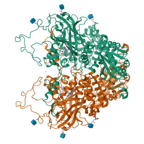
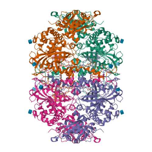
| CATALASE | A, B [auth E] | 688 | Penicillium janthinellum | Mutation(s): 0 EC: 1.11.1.6 | 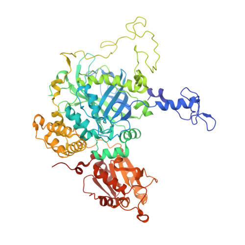 |
UniProt | |||||
Find proteins for D9N167 (Penicillium janthinellum) Explore D9N167 Go to UniProtKB: D9N167 | |||||
Entity Groups | |||||
| Sequence Clusters | 30% Identity50% Identity70% Identity90% Identity95% Identity100% Identity | ||||
| UniProt Group | D9N167 | ||||
Glycosylation | |||||
| Glycosylation Sites: 3 | |||||
Sequence AnnotationsExpand | |||||
| |||||
| Ligands 4 Unique | |||||
|---|---|---|---|---|---|
| ID | Chains | Name / Formula / InChI Key | 2D Diagram | 3D Interactions | |
| HDD Query on HDD | C [auth A], K [auth E] | CIS-HEME D HYDROXYCHLORIN GAMMA-SPIROLACTONE C34 H32 Fe N4 O5 UMGOPAWIGKFTRK-QQDQPIDJSA-N |  | ||
| NAG Query on NAG | D [auth A] E [auth A] F [auth A] L [auth E] M [auth E] | 2-acetamido-2-deoxy-beta-D-glucopyranose C8 H15 N O6 OVRNDRQMDRJTHS-FMDGEEDCSA-N |  | ||
| 3TR Query on 3TR | G [auth A], O [auth E] | 3-AMINO-1,2,4-TRIAZOLE C2 H4 N4 KLSJWNVTNUYHDU-UHFFFAOYSA-N |  | ||
| CA Query on CA | H [auth A] I [auth A] J [auth A] P [auth E] Q [auth E] | CALCIUM ION Ca BHPQYMZQTOCNFJ-UHFFFAOYSA-N |  | ||
| Length ( Å ) | Angle ( ˚ ) |
|---|---|
| a = 144.3 | α = 90 |
| b = 144.3 | β = 90 |
| c = 133.8 | γ = 120 |
| Software Name | Purpose |
|---|---|
| REFMAC | refinement |
| DENZO | data reduction |
| SCALEPACK | data scaling |
| MOLREP | phasing |
RCSB PDB is hosted by

RCSB PDB is a member of the