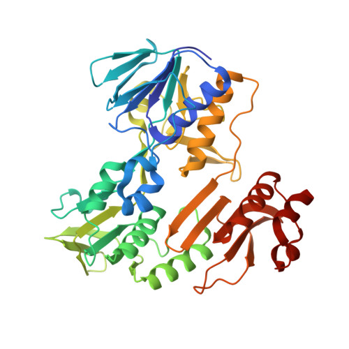
RCSB PDB - 2YVG: crystal structure of ferredoxin reductase, BPHA4 (blue-semiquinone)

 2YVG

crystal structure of ferredoxin reductase, BPHA4 (blue-semiquinone)


Experimental Data Snapshot

  • Method: X-RAY DIFFRACTION
  • Resolution: 1.60 Å
  • R-Value Free: 0.220 
  • R-Value Work: 0.194 
  • R-Value Observed: 0.195 

wwPDB Validation   3D Report Full Report


Ligand Structure Quality Assessment 

Created with Raphaël 2.3.0Worse 01 BetterLigand structure goodness of fit to experimental dataBest fitted FADClick on this verticalbar to view detailsBest fitted NADClick on this verticalbar to view details

This is version 1.2 of the entry. See complete history


Literature

Molecular Mechanism of the Redox-dependent Interaction between NADH-dependent Ferredoxin Reductase and Rieske-type [2Fe-2S] Ferredoxin

Senda, M.Kishigami, S.Kimura, S.Fukuda, M.Ishida, T.Senda, T.

(2007) J Mol Biol 373: 382-400

  • DOI: https://doi.org/10.1016/j.jmb.2007.08.002
  • Primary Citation of Related Structures:  
    2E4P, 2E4Q, 2GQW, 2GR0, 2YVF, 2YVG, 2YVJ

  • PubMed Abstract: 

    The electron transfer system of the biphenyl dioxygenase BphA, which is derived from Acidovorax sp. (formally Pseudomonas sp.) strain KKS102, is composed of an FAD-containing NADH-ferredoxin reductase (BphA4) and a Rieske-type [2Fe-2S] ferredoxin (BphA3). Biochemical studies have suggested that the whole electron transfer process from NADH to BphA3 comprises three consecutive elementary electron-transfer reactions, in which BphA3 and BphA4 interact transiently in a redox-dependent manner. Initially, BphA4 receives two electrons from NADH. The reduced BphA4 then delivers one electron each to the [2Fe-2S] cluster of the two BphA3 molecules through redox-dependent transient interactions. The reduced BphA3 transports the electron to BphA1A2, a terminal oxygenase, to support the activation of dioxygen for biphenyl dihydroxylation. In order to elucidate the molecular mechanisms of the sequential reaction and the redox-dependent interaction between BphA3 and BphA4, we determined the crystal structures of the productive BphA3-BphA4 complex, and of free BphA3 and BphA4 in all the redox states occurring in the catalytic cycle. The crystal structures of these reaction intermediates demonstrated that each elementary electron transfer induces a series of redox-dependent conformational changes in BphA3 and BphA4, which regulate the interaction between them. In addition, the conformational changes induced by the preceding electron transfer seem to induce the next electron transfer. The interplay of electron transfer and induced conformational changes seems to be critical to the sequential electron-transfer reaction from NADH to BphA3.


  • Organizational Affiliation

    Japan Biological Information Research Center, Japan Biological Informatics Consortium, 2-42 Aomi, Koto-ku, Tokyo 135-0064, Japan.


Macromolecules
Find similar proteins by:  (by identity cutoff)  |  3D Structure
Entity ID: 1
MoleculeChains Sequence LengthOrganismDetailsImage
Ferredoxin reductase408Pseudomonas sp.Mutation(s): 0 
Gene Names: bphA4
EC: 1.18.1.2
UniProt
Find proteins for Q52437 (Pseudomonas sp. (strain KKS102))
Explore Q52437 
Go to UniProtKB:  Q52437
Entity Groups  
Sequence Clusters30% Identity50% Identity70% Identity90% Identity95% Identity100% Identity
UniProt GroupQ52437
Sequence Annotations
Expand
  • Reference Sequence
Small Molecules
Ligands 2 Unique
IDChains Name / Formula / InChI Key2D Diagram3D Interactions
FAD
Query on FAD

Download Ideal Coordinates CCD File 
B [auth A]FLAVIN-ADENINE DINUCLEOTIDE
C27 H33 N9 O15 P2
VWWQXMAJTJZDQX-UYBVJOGSSA-N
NAD
Query on NAD

Download Ideal Coordinates CCD File 
C [auth A]NICOTINAMIDE-ADENINE-DINUCLEOTIDE
C21 H27 N7 O14 P2
BAWFJGJZGIEFAR-NNYOXOHSSA-N
Experimental Data & Validation

Experimental Data

  • Method: X-RAY DIFFRACTION
  • Resolution: 1.60 Å
  • R-Value Free: 0.220 
  • R-Value Work: 0.194 
  • R-Value Observed: 0.195 
  • Space Group: P 61 2 2
Unit Cell:
Length ( Å )Angle ( ˚ )
a = 98.288α = 90
b = 98.288β = 90
c = 170.066γ = 120
Software Package:
Software NamePurpose
XTALVIEWrefinement
REFMACrefinement
ADSCdata collection
XDSdata scaling

Structure Validation

View Full Validation Report


Ligand Structure Quality Assessment 

Created with Raphaël 2.3.0Worse 01 BetterLigand structure goodness of fit to experimental dataBest fitted FADClick on this verticalbar to view detailsBest fitted NADClick on this verticalbar to view details

Entry History 

Deposition Data

Revision History  (Full details and data files)

  • Version 1.0: 2007-10-16
    Type: Initial release
  • Version 1.1: 2011-07-13
    Changes: Advisory, Version format compliance
  • Version 1.2: 2024-03-13
    Changes: Data collection, Database references, Derived calculations