2ZK9 | pdb_00002zk9

Crystal Structure of Protein-glutaminase


Experimental Data Snapshot

  • Method: X-RAY DIFFRACTION
  • Resolution: 1.15 Å
  • R-Value Free: 
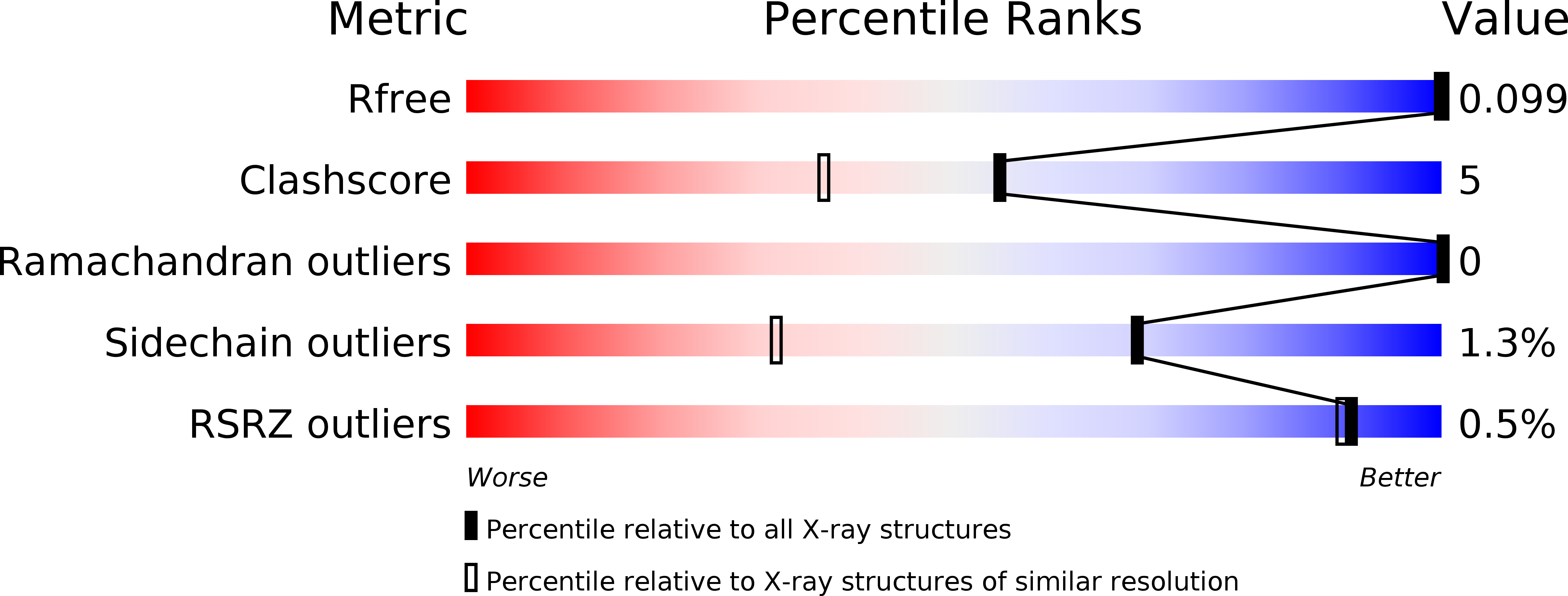
    0.136 (Depositor), 0.097 (DCC) 
  • R-Value Work: 
    0.098 (Depositor), 0.100 (DCC) 

wwPDB Validation   3D Report Full Report


This is version 1.3 of the entry. See complete history


Literature

Crystal structures of protein glutaminase and its pro forms converted into enzyme-substrate complex

Hashizume, R.Maki, Y.Mizutani, K.Takahashi, N.Matsubara, H.Sugita, A.Sato, K.Yamaguchi, S.Mikami, B.

(2011) J Biological Chem 286: 38691-38702

  • DOI: https://doi.org/10.1074/jbc.M111.255133
  • Primary Citation of Related Structures:  
    2ZK9, 3A54, 3A55, 3A56

  • PubMed Abstract: 

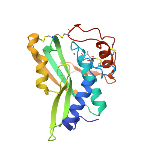
    Protein glutaminase, which converts a protein glutamine residue to a glutamate residue, is expected to be useful as a new food-processing enzyme. The crystal structures of the mature and pro forms of the enzyme were refined at 1.15 and 1.73 Å resolution, respectively. The overall structure of the mature enzyme has a weak homology to the core domain of human transglutaminase-2. The catalytic triad (Cys-His-Asp) common to transglutaminases and cysteine proteases is located in the bottom of the active site pocket. The structure of the recombinant pro form shows that a short loop between S2 and S3 in the proregion covers and interacts with the active site of the mature region, mimicking the protein substrate of the enzyme. Ala-47 is located just above the pocket of the active site. Two mutant structures (A47Q-1 and A47Q-2) refined at 1.5 Å resolution were found to correspond to the enzyme-substrate complex and an S-acyl intermediate. Based on these structures, the catalytic mechanism of protein glutaminase is proposed.


  • Organizational Affiliation

    Laboratory of Applied Structural Biology, Division of Applied Life Sciences, Graduate School of Agriculture, Kyoto University, Gokasho, Uji, Kyoto 611-0011, Japan.


Macromolecules
Find similar proteins by:  (by identity cutoff)  |  3D Structure
Entity ID: 1
MoleculeChains Sequence LengthOrganismDetailsImage
Protein-glutaminaseA [auth X]185Chryseobacterium proteolyticumMutation(s): 0 
EC: 3.5.1
UniProt
Find proteins for Q9AQQ8 (Chryseobacterium proteolyticum)
Explore Q9AQQ8 
Go to UniProtKB:  Q9AQQ8
Entity Groups  
Sequence Clusters30% Identity50% Identity70% Identity90% Identity95% Identity100% Identity
UniProt GroupQ9AQQ8
Sequence Annotations
Expand
  • Reference Sequence
Experimental Data & Validation

Experimental Data

  • Method: X-RAY DIFFRACTION
  • Resolution: 1.15 Å
  • R-Value Free:  0.136 (Depositor), 0.097 (DCC) 
  • R-Value Work:  0.098 (Depositor), 0.100 (DCC) 
Space Group: P 65 2 2
Unit Cell:
Length ( Å )Angle ( ˚ )
a = 62.306α = 90
b = 62.306β = 90
c = 185.532γ = 120
Software Package:
Software NamePurpose
SHELXmodel building
SHELXL-97refinement
SHELXphasing

Structure Validation

View Full Validation Report


Entry History 

Deposition Data

  • Released Date: 2009-03-17 
  • Deposition Author(s): Hashizume, R.
  • This entry supersedes: 2Z8T

Revision History  (Full details and data files)

  • Version 1.0: 2009-03-17
    Type: Initial release
  • Version 1.1: 2011-07-13
    Changes: Non-polymer description, Version format compliance
  • Version 1.2: 2012-08-29
    Changes: Database references, Other
  • Version 1.3: 2024-11-20
    Changes: Data collection, Database references, Derived calculations, Structure summary