2ZQO | pdb_00002zqo

Crystal structure of the earthworm R-type lectin C-half in complex with GalNAc


Experimental Data Snapshot

  • Method: X-RAY DIFFRACTION
  • Resolution: 1.80 Å
  • R-Value Free: 
    0.260 (Depositor), 0.260 (DCC) 
  • R-Value Work: 
    0.192 (Depositor), 0.190 (DCC) 
  • R-Value Observed: 
    0.195 (Depositor) 

Starting Model: experimental
View more details

wwPDB Validation   3D Report Full Report


Ligand Structure Quality Assessment 

Created with Raphaël 2.3.0Worse 01 BetterLigand structure goodness of fit to experimental dataBest fitted NGAClick on this verticalbar to view details

This is version 1.3 of the entry. See complete history


Literature

Sugar-complex structures of the C-half domain of the galactose-binding lectin EW29 from the earthworm Lumbricus terrestris

Suzuki, R.Kuno, A.Hasegawa, T.Hirabayashi, J.Kasai, K.Momma, M.Fujimoto, Z.

(2009) Acta Crystallogr D Biol Crystallogr 65: 49-57

  • DOI: https://doi.org/10.1107/S0907444908037451
  • Primary Citation of Related Structures:  
    2ZQN, 2ZQO

  • PubMed Abstract: 

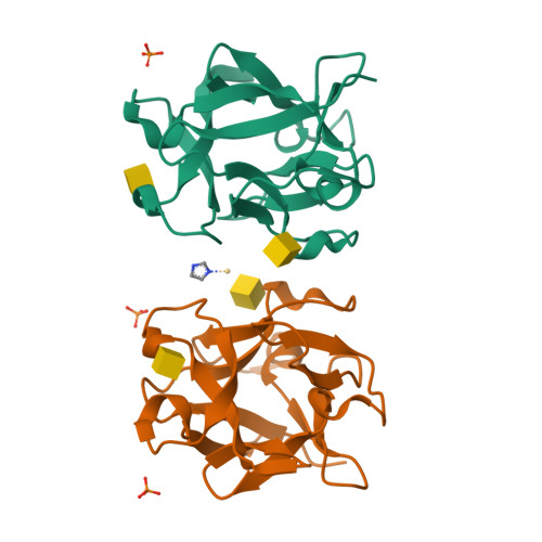
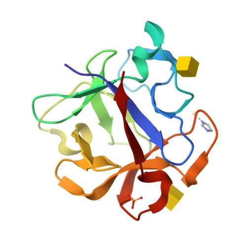
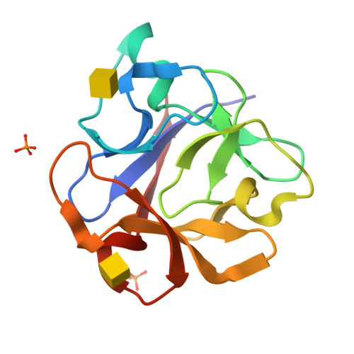
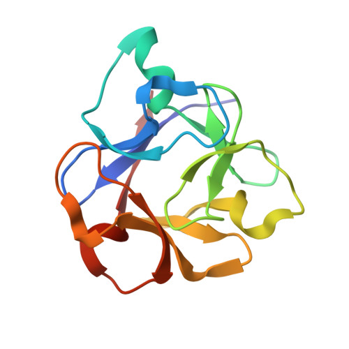
    R-type lectins are one of the most prominent types of lectin; they exist ubiquitously in nature and mainly bind to the galactose unit of sugar chains. The galactose-binding lectin EW29 from the earthworm Lumbricus terrestris belongs to the R-type lectin family as represented by the plant lectin ricin. It shows haemagglutination activity and is composed of a single peptide chain that includes two homologous domains: N-terminal and C-terminal domains. A truncated mutant of EW29 comprising the C-terminal domain (rC-half) has haemagglutination activity by itself. In order to clarify how rC-half recognizes ligands and shows haemagglutination activity, X-ray crystal structures of rC-half in complex with D-lactose and N-acetyl-D-galactosamine have been determined. The structure of rC-half is similar to that of the ricin B chain and consists of a beta-trefoil fold; the fold is further divided into three similar subdomains referred to as subdomains alpha, beta and gamma, which are gathered around the pseudo-threefold axis. The structures of sugar complexes demonstrated that subdomains alpha and gamma of rC-half bind terminal galactosyl and N-acetylgalactosaminyl glycans. The sugar-binding properties are common to both ligands in both subdomains and are quite similar to those of ricin B chain-lactose complexes. These results indicate that the C-terminal domain of EW29 uses these two galactose-binding sites for its function as a single-domain-type haemagglutinin.


  • Organizational Affiliation

    Protein Research Unit, National Institute of Agrobiological Sciences, Tsukuba 305-8602, Japan.


Macromolecules
Find similar proteins by:  (by identity cutoff)  |  3D Structure
Entity ID: 1
MoleculeChains Sequence LengthOrganismDetailsImage
29-kDa galactose-binding lectin
A, B
130Lumbricus terrestrisMutation(s): 0 
UniProt
Find proteins for O96048 (Lumbricus terrestris)
Explore O96048 
Go to UniProtKB:  O96048
Entity Groups  
Sequence Clusters30% Identity50% Identity70% Identity90% Identity95% Identity100% Identity
UniProt GroupO96048
Sequence Annotations
Expand
  • Reference Sequence
Small Molecules
Ligands 4 Unique
IDChains Name / Formula / InChI Key2D Diagram3D Interactions
NGA
Query on NGA

Download Ideal Coordinates CCD File 
C [auth A],
D [auth A],
H [auth B],
I [auth B]
2-acetamido-2-deoxy-beta-D-galactopyranose
C8 H15 N O6
OVRNDRQMDRJTHS-JAJWTYFOSA-N
CD
Query on CD

Download Ideal Coordinates CCD File 
E [auth A]CADMIUM ION
Cd
WLZRMCYVCSSEQC-UHFFFAOYSA-N
PO4
Query on PO4

Download Ideal Coordinates CCD File 
F [auth A],
J [auth B],
K [auth B]
PHOSPHATE ION
O4 P
NBIIXXVUZAFLBC-UHFFFAOYSA-K
IMD
Query on IMD

Download Ideal Coordinates CCD File 
G [auth A]IMIDAZOLE
C3 H5 N2
RAXXELZNTBOGNW-UHFFFAOYSA-O
Experimental Data & Validation

Experimental Data

  • Method: X-RAY DIFFRACTION
  • Resolution: 1.80 Å
  • R-Value Free:  0.260 (Depositor), 0.260 (DCC) 
  • R-Value Work:  0.192 (Depositor), 0.190 (DCC) 
  • R-Value Observed: 0.195 (Depositor) 
Space Group: C 1 2 1
Unit Cell:
Length ( Å )Angle ( ˚ )
a = 123.21α = 90
b = 34.747β = 104.2
c = 61.586γ = 90
Software Package:
Software NamePurpose
REFMACrefinement
HKL-2000data collection
DENZOdata reduction
SCALEPACKdata scaling
MOLREPphasing

Structure Validation

View Full Validation Report


Ligand Structure Quality Assessment 

Created with Raphaël 2.3.0Worse 01 BetterLigand structure goodness of fit to experimental dataBest fitted NGAClick on this verticalbar to view details

Entry History 

Deposition Data

Revision History  (Full details and data files)

  • Version 1.0: 2008-09-02
    Type: Initial release
  • Version 1.1: 2011-07-13
    Changes: Version format compliance
  • Version 1.2: 2020-07-29
    Type: Remediation
    Reason: Carbohydrate remediation
    Changes: Data collection, Derived calculations, Structure summary
  • Version 1.3: 2023-11-01
    Changes: Data collection, Database references, Refinement description, Structure summary